TOM
-
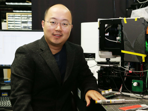 Meet the KAISTian of 2017, Professor YongKeun Park
Professor YongKeun Park from the Department of Physics is one of the star professors in KAIST. Rising to the academic stardom, Professor Park’s daily schedule is filled with series of business meetings in addition to lab meetings and lectures.
The year 2017 must have been special for him. During the year, he published numerous papers in international journals, such as Nature Photonics, Nature Communications and Science Advances. These high performances drew international attention from renowned media, including Newsweek and Forbes. Moreover, recognizing his research performance, he was elected as a fellow member of the Optical Society (OSA) in his mid-30s. Noting that the members’ age ranges from late 50s to early 60s, Professor Park’s case considered to be quite exceptional.
Adding to his academic achievement, he has launched two startups powered of his own technologies. One is called Tomocube, a company specialized in 3-D imaging microscope using holotomography technology. His company is currently exporting the products to multiple countries, including the United States and Japan. The other one is The.Wave.Talk which has technologies for examining pre-existing bacteria anywhere and anytime.
His research career and entrepreneurship are well deserved recipient of many honors. At the 2018 kick-off ceremony, Professor Park was awarded the KAISTian of 2017 in recognition of his developing holographic measure and control technology as well as founding a new field for technology application.
KAISTian of the Year, first presented in 2001, is an award to recognize the achievements and exemplary contribution of KAIST member who has put significant effort nationally and internationally, enhancing the value of KAIST.
While receiving the award, he thanked his colleagues and his students who have achieved this far together. He said, “I would like to thank KAIST for providing environment for young professors like me so that we can engage themselves in research. Also, I would like to mention that I am an idea seeder and my students do the most of the research. So, I appreciate my students for their hard works, and it is very pleasure to have them. Lastly, I thank the professors for teaching these outstanding students. I feel great responsibility over this title. I will dedicate myself to make further progress in commercializing technology in KAIST.”
Expecting his successful startup cases as a model and great inspiration to students as well as professors, KAIST interviewed Professor Park.
Q What made you decide to found your startups?
A I believed that my research areas could be further used. As a professor, I believe that it is a university’s role to create added value through commercializing technology and creating startups.
Q You have co-founded two startups. What is your role in each company?
A So, basically I have two full-time jobs, professor in KAIST and CTO in Tomocube. After transferring the technology, I hold the position of advisor in The.Wave.Talk.
(Holographic images captured by the product Professor Park developed)
Q Do your students also participate in your companies or can they?
A No, the school and companies are separate spaces; in other words, they are not participating in my companies. They have trained my employees when transferring the technologies, but they are not directly working for the companies.
However, they can participate if they want to. If there’s a need to develop a certain technology, an industry-academia contract can be made. According to the agreement, students can work for the companies.
Q Were there any hardships when preparing the startups?
A At the initial stage, I did not have a financial problem, thanks to support from Startup KAIST. Yet, inviting capital is the beginning, and I think every step I made to operate, generate revenue, and so on is not easy.
Q Do you believe KAIST is startup-friendly?
A Yes, there’s no school like KAIST in Korea and any other country. Besides various programs to support startup activities, Startup KAIST has many professors equipped with a great deal of experience. Therefore, I believe that KAIST provides an excellent environment for both students and professors to create startups.
Q Do you have any suggestion to KAIST institutionally?
A Well, I would like to make a comment to students and professors in KAIST. I strongly recommend them to challenge themselves by launching startups if they have good ideas. Many students wish to begin their jobs in government-funded research institutes or major corporates, but I believe that engaging in a startup company will also give them valuable and very productive experience.
Unlike before, startup institutions are well established, so attracting good capital is not so hard. There are various activities offered by Startup KAIST, so it’s worthwhile giving it a try.
Q What is your goal for 2018 as a professor and entrepreneur?
A I don’t have a grand plan, but I will work harder to produce good students with new topics in KAIST while adding power to my companies to grow bigger.
By Se Yi Kim from the PR Office
2018.01.03 View 13293
 -
 Strengthening Industry-Academia Cooperation with LG CNS
On November 20, KAIST signed an MoU with LG CNS for industry-academia partnership in education, research, and business in the fields of AI and Big Data. Rather than simply developing education programs or supporting industry-academia scholarships, both organizations agreed to carry out a joint research project on AI and Big Data that can be applied to practical business.
KAIST will collaborate with LG CNS in the fields of smart factories, customer analysis, and supply chain management analysis.
Not only will LG CNS offer internships to KAIST students, but it also will support professors and students who propose innovative startup ideas for AI and Big Data. Offering an industry-academia scholarship for graduate students is also being discussed. Together with LG CNS, KAIST will put its efforts into propose projects regarding AI and Big Data in the public sector.
Furthermore, KAIST and LG CNS will jointly explore and carry out industry-academia projects that could be practically used in business. Both will carry out the project vigorously through strong cooperation; for instance, LG CNS employees can be assigned to KAIST, if necessary. Also, LG CNS’s AI and Big Data platform, called DAP (Data Analytics & AI Platform) will be used as a data analysis tool during the project and the joint outcomes will be installed in DAP.
KAIST professors with expertise in AI deep learning have trained LG CNS employees since the Department of Industrial & Systems Engineering established ‘KAIST AI Academy’ in LG CNS last August.
“With KAIST, the best research-centered university in Korea, we will continue to lead in developing the field of AI and Big Data and provide innovative services that create value by connecting them to customer business,” Yong Shub Kim, the CEO of LG CNS, highlighted.
2017.11.22 View 13167
 -
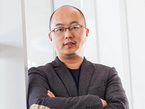 Professor YongKeun Park Elected as a Fellow of the Optical Society
Professor YongKeun Park, from the Department of Physics at KAIST, was elected as a fellow member of the Optical Society (OSA) in Washington, D.C. on September 12. Fellow membership is given to members who have made a significant contribution to the advancement of optics and photonics.
Professor Park was recognized for his research on digital holography and wavefront control technology.
Professor Park has been producing outstanding research outcomes in the field of holographic technology and light scattering control since joining KAIST in 2010. In particular, he developed and commercialized technology for a holographic telescope. He applied it to various medical and biological research projects, leading the field worldwide.
In the past, cells needed to be dyed with fluorescent materials to capture a 3-D image. However, Professor Park’s holotomography (HT) technology can capture 3-D images of living cells and tissues in real time without color dyeing. This technology allows diversified research in the biological and medical field.
Professor Park established a company, Tomocube, Inc. in 2015 to commercialize the technology. In 2016, he received funding from SoftBank Ventures and Hanmi Pharmaceutical. Currently, major institutes, including MIT, the University of Pittsburgh, the German Cancer Research Center, and Seoul National University Hospital are using his equipment.
Recently, Professor Park and his team developed technology based on light scattering measurements. With this technology, they established a company called The Wave Talk and received funding from various organizations, such as NAVER. Its first product is about to be released.
Professor Park said, “I am glad to become a fellow member based on the research outcomes I produced since I was appointed as a professor at KAIST. I would like to thank the excellent researchers as well as the school for its support. I will devote myself to continuously producing novel outcomes in both basic and applied fields.”
Professor Park has published nearly 100 papers in renowned journals including Nature Photonics, Nature Communications, Science Advances, and Physical Review Letters.
2017.10.18 View 13913
 -
 Professor Kwon to Represent the Asia-Pacific Region of the IEEE RAS
Professor Dong-Soon Kwon of the Mechanical Engineering Department at KAIST has been reappointed to the Administrative Committee of the Institute of Electrical and Electronics Engineers (IEEE) Robotics and Automation Society (IEEE RAS). Beginning January 1, 2017, he will serve his second three-year term, which will end in 2019. In 2014, he was the first Korean appointed to the committee, representing the Asia-Pacific community of the IEEE Society.
Professor Kwon said, “I feel thankful but, at the same time, it is a great responsibility to serve the Asian research community within the Society. I hope I can contribute to the development of robotics engineering in the region and in Korea as well.”
Consisted of 18 elected members, the administrative committee manages the major activities of IEEE RAS including hosting its annual flagship meeting, the International Conference on Robotics and Automation.
The IEEE RAS fosters the advancement in the theory and practice of robotics and automation engineering and facilitates the exchange of scientific and technological knowledge that supports the maintenance of high professional standards among its members.
2016.12.06 View 11476
 -
 Professor Tae-Eog Lee Receives December's Scientist of the Month Award by the Korean Government
Professor Tae-Eog Lee of the Industrial and Systems Engineering at KAIST received the Scientist of the Month Award for December 2015. The award is sponsored by the Ministry of Science, ICT and Future Planning of Korea, which was hosted by the National Research Foundation of Korea.
The award recognizes Professor Lee’s efforts to advance the field of semiconductor device fabrication processing. This includes the development of the most efficient scheduling and controlling of cluster tools. He also created mathematical solutions to optimize the complicated cycle time of cluster tools in semiconductor manufacturing and the process of robot task workload.
Professor Lee contributed to the formation of various discrete event systems and automation systems based on his mathematical theories and solutions and advanced a scheduling technology for the automation of semiconductor production.
He has published 18 research papers in the past three years and has pioneered to develop Korean tool schedulers through the private sector-university cooperation.
2015.12.10 View 8912
 -
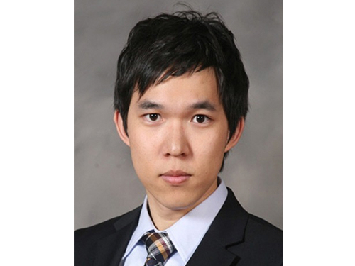 Dong-Young Lee, a Doctoral Candidate, Receives the Best Paper Award
Dong-Young Lee, a Ph.D. candidate in the Mechanical Engineering Department, KAIST, received the Best Paper Award at the 18th International Conference on Composite Structures (ICCS). The event was held in Lisbon, Portugal, on June 15-18, 2015. Mr. Lee’s adviser is Professor Dai-Gil Lee of the same department.
The ICCS is held every other year, and is one of the largest and long-established conferences on composite materials and structures in the world. At this year’s conference, a total of 680 papers were presented, among which, two papers were chosen for the Best Paper Award, including Mr. Lee’s.
The paper, entitled “Gasket-integrated Carbon and Silicon Elastomer Composite Bipolar Plate for High-temperature PEMFC,” will be published in the September issue of Composite Structures which is one of the top journals in mechanical engineering as judged by the Google Scholar Metrics rankings.
Mr. Lee dropped the conventional method of PEMFC (Proton Exchange Membrane Fuel Cells) assembly and instead developed a gasket-integrated carbon and silicone elastomer composite bipolar plate. This technology significantly increased the energy efficiency of fuel cells and their productivity.
Mr. Lee said, “I would like to thank the many people who supported me, especially my Ph.D. adviser, Professor Dai-Gil Lee. Without their encouragement, I would have not won this award. I hope my research will contribute to solving energy problems in the future.”
In addition, Professor Joon-Woo Im from Chonbuk National University, Senior Researcher Il-Bum Choi from the Agency for Defense Development, and a fellow doctoral candidate Soo-Hyun Nam from KAIST participated in this research project.
2015.07.09 View 11299
 -
 Professor Naehyuck Jang was Appointed Technical Program Chair of the Design Automation Conference
Professor Naehyuck Jang of the Electrical Engineering Department at KAIST was appointed as the technical program chair of the Design Automation Conference (DAC). He is the first Asian to serve the conference as the chair. At next year’s conference, he will select 150 program committee members and supervise the selection process of 1,000 papers.
Founded in 1964, DAC encompasses research related to automation of semiconductor processes, which usually involve billions of transistors. More than seven thousand people and 150 companies from all around the world participate, of which only the top 20% of the submitted papers are selected. It is the most prestigious conference in the field of semiconductor automation.
The Design Automation Conference also introduces optimization and automation of design processes of systems, hardware security, automobiles, and the Internet of things. Professor Jang specializes in low power system designs. As an ACM Distinguished Scientist, Professor Jang was elected as the chairman after contributing to this year’s program committee by reforming the process of selection of papers.
Professor Jang said, “This year’s conference represents a departure, where we move from the field of traditional semiconductors to the optimization of embedded system, the Internet of things, and security. He added that “we want to create a paper selection process that can propose the future of design automation.”
The 53rd annual DAC will take place at the Austin Convention Center in Texas in June 2016.
2015.07.02 View 8058
 -
 Professor Rim Presents at IAEA Workshop in Vienna
Professor Chun-Taek Rim of the Department of Nuclear and Quantum Engineering at KAIST recently attended the International Atomic Energy Agency (IAEA)’s workshop on the Application of Wireless Technologies in Nuclear Power Plant Instrumentation and Control System. It took place on March 30-April 2, 2015, in Vienna, Austria.
Representing Korea, Professor Rim gave a talk entitled “Highly Reliable Wireless Power and Communications under Severe Accident of Nuclear Power Plants (NPPs).” About 20 industry experts from 12 countries such as AREVA (France), Westinghouse (US), Oak Ridge National Laboratory (US), Hitachi (Japan), and ENEA (Italy) joined the meeting.
The IAEA hosted the workshop to explore the application of wireless technology for the operation and management of NPPs. It formed a committee consisting of eminent professionals worldwide in NPP instrumentation and control systems, communications, and nuclear power to examine this issue in-depth and to conduct various research projects for the next three years.
In particular, the committee will concentrate its research on improving the reliability and safety of using wireless technology, not only in the normal operation of nuclear plants but also in extreme conditions such as the Fukushima Daiichi nuclear accident. The complementation, economic feasibility, and standardization of NPPs when applying wireless technology will be also discussed.
Professor Rim currently leads the Nuclear Power Electronics
and Robotics Lab at KAIST (http://tesla.kaist.ac.kr/index_eng.php?lag=eng).
Picture 1: Professors Rim presents his topic at the IAEA Workshop in Vienna.
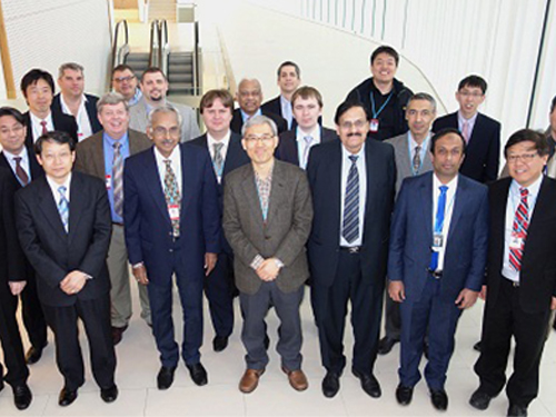
Picture 2: The IAEA Workshop Participants
2015.04.07 View 14208
 -
 System Approach Using Metabolite Structural Similarity Toward TOM Suggested
A Korean research team at KAIST suggests that a system approach using metabolite structural similarity helps to elucidate the mechanisms of action of traditional oriental medicine.
Traditional oriental medicine (TOM) has been practiced in Asian countries for centuries, and is gaining increasing popularity around the world. Despite its efficacy in various symptoms, TOM has been practiced without precise knowledge of its mechanisms of action. Use of TOM largely comes from empirical knowledge practiced over a long period of time. The fact that some of the compounds found in TOM have led to successful modern drugs such as artemisinin for malaria and taxol (Paclitaxel) for cancer has spurred modernization of TOM.
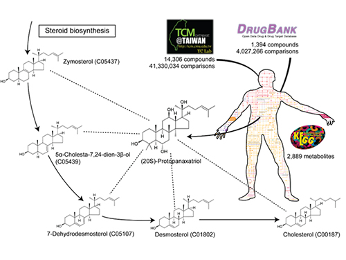
A research team led by Sang-Yup Lee at KAIST has focused on structural similarities between compounds in TOM and human metabolites to help explain TOM’s mechanisms of action. This systems approach using structural similarities assumes that compounds which are structurally similar to metabolites could affect relevant metabolic pathways and reactions by biosynthesizing structurally similar metabolites.
Structural similarity analysis has helped to identify mechanisms of action of TOM. This is described in a recent study entitled “A systems approach to traditional oriental medicine,” published online in Nature Biotechnology on March 6, 2015. In this study, the research team conducted structural comparisons of all the structurally known compounds in TOM and human metabolites on a large-scale. As a control, structures of all available approved drugs were also compared against human metabolites. This structural analysis provides two important results. First, the identification of metabolites structurally similar to TOM compounds helped to narrow down the candidate target pathways and reactions for the effects from TOM compounds. Second, it suggested that a greater fraction of all the structurally known TOM compounds appeared to be more similar to human metabolites than the approved drugs. This second finding indicates that TOM has a great potential to interact with diverse metabolic pathways with strong efficacy. This finding, in fact, shows that TOM compounds might be advantageous for the multitargeting required to cure complex diseases. “Once we have narrowed down candidate target pathways and reactions using this structural similarity approach, additional in silico tools will be necessary to characterize the mechanisms of action of many TOM compounds at a molecular level,” said Hyun Uk Kim, a research professor at KAIST.
TOM’s multicomponent, multitarget approach wherein multiple components show synergistic effects to treat symptoms is highly distinctive. The researchers investigated previously observed effects recorded since 2000 of a set of TOM compounds with known mechanisms of action. TOM compounds’ synergistic combinations largely consist of a major compound providing the intended efficacy to the target site and supporting compounds which maximize the efficacy of the major compound. In fact, such combination designs appear to mirror the Kun-Shin-Choa-Sa design principle of TOM.
That principle, Kun-Shin-Choa-Sa (君臣佐使 or Jun-Chen-Zuo-Shi in Chinese) literally means “king-minister-assistant-ambassador.” In ancient East Asian medicine, treating human diseases and taking good care of the human body are analogous to the politics of governing a nation. Just as good governance requires that a king be supported by ministers, assistants and/or ambassadors, treating diseases or good care of the body required the combined use of herbal medicines designed based on the concept of Kun-Shin-Choa-Sa. Here, the Kun (king or the major component) indicates the major medicine (or herb) conveying the major drug efficacy, and is supported by three different types of medicines: the Shin (minister or the complementary component) for enhancing and/or complementing the efficacy of the Kun, Choa (assistant or the neutralizing component) for reducing any side effects caused by the Kun and reducing the minor symptoms accompanying major symptom, and Sa (ambassador or the delivery/retaining component) which facilitated the delivery of the Kun to the target site, and retaining the Kun for prolonged availability in the cells.
The synergistic combinations of TOM compounds reported in the literature showed four different types of synergisms: complementary action (similar to Kun-Shin), neutralizing action (similar to Kun-Choa), facilitating action or pharmacokinetic potentiation (both similar to Kun-Choa or Kun-Sa). Additional structural analyses for these compounds with synergism show that they appeared to affect metabolism of amino acids, co-factors and vitamins as major targets.
Professor Sang Yup Lee remarks, “This study lays a foundation for the integration of traditional oriental medicine with modern drug discovery and development. The systems approach taken in this analysis will be used to elucidate mechanisms of action of unknown TOM compounds which will then be subjected to rigorous validation through clinical and in silico experiments.”
Sources: Kim, H.U. et al. “A systems approach to traditional oriental medicine.” Nature Biotechnology. 33: 264-268 (2015).
This work was supported by the Bio-Synergy Research Project (2012M3A9C4048759) of the Ministry of Science, ICT and Future Planning through the National Research Foundation. This work was also supported by the Novo Nordisk Foundation.
The picture below presents the structural similarity analysis of comparing compounds in traditional oriental medicine and those in all available approved drugs against the structures of human metabolites.
2015.03.09 View 11490
 -
 Professor Joong-Keun Park Receives SeAH Heam Academic Award
Professor Joong-Keun Park of the Department of Materials Science and Engineering at KAIST received an award from SeAH Steel Corp. in recognition of his academic achievements in the field of metallic and materials engineering.
The award was presented at the 2014 Fall Conference of the Korean Institute of Metals and Materials which took place on October 22-24 at the Kangwon Land Convention Hotel.
The award, called “SeAH Heam Academic Award,” is given annually to a scholar who has contributed to the development of new metal and polymer composite materials and its related field in Korea. Following the award ceremony, Professor Park gave a keynote speech on ferrous metals for automotive materials.
2014.11.04 View 8920
 -
 First Instance of Negative Effects from Terahertz-Range Electromagnetic Waves
Professor Philhan Kim
Electromagnetic waves (EM-wave) in the terahertz range were widely regarded as the “dream wavelength” due to its perceived neutrality. Its application was also wider than X-rays. However, KAIST scientists have discovered negative effects from terahertz EM-waves.
Professor Philhan Kim of KAIST’s Graduate School of Nanoscience and Technology and Dr. Young-wook Jeong of the Korea Atomic Energy Research Institute (KAERI) observed inflammation of animal skin tissue when exposed to terahertz EM-waves.
The results were published in the online edition of Optics Express (May 19, 20104).
Terahertz waves range from 0.1 to 10 terahertz and have a longer wavelength than visible or infrared light. Commonly used to see through objects like the X-ray, it was believed that the low energy of terahertz waves did not inflict any harm on the human body.
Despite being applied for security checks, next-generation wireless communications, and medical imaging technology, little research has been conducted in proving its safety and impact. Conventional research failed to predict the exact impact of terahertz waves on organic tissues as only artificially cultured cells were used.
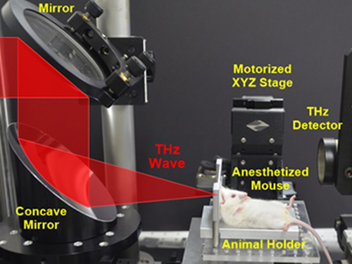
The research team at KAERI developed a high power terahertz EM-wave generator that can be used on live organisms. A high power generator was necessary in applications such as biosensors and required up to 10 times greater power than currently used telecommunications EM-wave. Simultaneously, a KAIST research team developed a high speed, high resolution video-laser microscope that can distinguish cells within the organism.
The experiment exposed 30 minutes of terahertz EM-wave on genetically modified mice and found six times the normal number of inflammation cells in the skin tissue after six hours. It was the first instance where negative side effects of terahertz EM-wave were observed.
Professor Kim commented that “the research has set a standard for how we can use the terahertz EM-wave safely” and that “we will use this research to analyze and understand the effects of other EM-waves on organisms.”
2014.06.20 View 10791
 -
 An Exploratory Study on Smartphone Abuse among College Students
Professor Uichin Lee
Professor Uichin Lee of the Department of Knowledge Service Engineering, KAIST, and his research team developed a system that automatically diagnoses the levels of smartphone addiction based on an analysis of smartphone use records.
Professor Lee investigated the usage patterns of 95 smartphone users (college students) by conducting surveys and interviews and collecting logged data. The research team divided participants into “risk” and “non-risk” groups based on a self-reported rating scale to evaluate their abuse of smartphones. As a result, 36 students were categorized as “high risk” and 59 were categorized as “low risk.”
The researchers collected over 50,000 hours of smartphone use encompassing power levels, screen, battery status, application use, internet use, calling, and texting.
The results showed that the “high risk” group used only 1~2 applications, focusing on mobile messengers (Kakotalk, etc.) and SNS (Facebook, etc.).
In addition, a relationship was found between alarm function and addiction levels. Users who set alarms for Kakaotalk messages and SNS comments used smartphones for an additional 38 minutes per day on average.
Results also showed that “high risk” students were on their smartphones for 4 hours and 13 minutes per day, 46 minutes longer than “low risk” students who used smartphones for 3 hours and 27 minutes. The difference was prevalent during 6 am and noon, and 6pm and midnight. In addition, “high risk” students accessed their smartphones 11.4 times more than “low risk” students.
Based on the collected data, Professor Lee developed an automatic system that distinguished users into “high risk” or “low risk” categories with 80% accuracy. The new system is expected to give an early diagnosis of addiction to smartphone users, thereby allowing for early treatment and intervention before the user becomes addicted.
Professor Lee commented that, "the conventional addiction analysis based on self-analysis surveys did not provide real-time data and were largely inaccurate. The new system overcomes these limitations through data science and personal big data analysis" and that he is "developing an application that monitors smartphone abuse."
Figure 1. Usage amount: overall and application-specific results
Figure 2. Usage frequency: overall and application-specific results
Figure 3. Overall diurnal usage time and frequency
2014.06.05 View 8418