ES
-
 A Study Reveals What Triggers Lung Damage during COVID-19
A longitudinal study of macrophages from SARS-CoV-2 infected lungs offers new insights into dynamic immunological changes
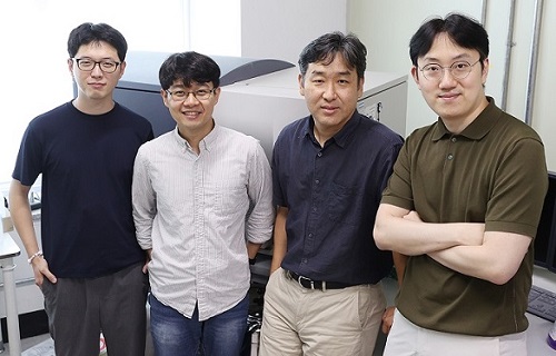
A KAIST immunology research team found that a specific subtype of macrophages that originated from blood monocytes plays a key role in the hyper-inflammatory response in SARS-CoV-2 infected lungs, by performing single-cell RNA sequencing of bronchoalveolar lavage fluid cells. This study provides new insights for understanding dynamic changes in immune responses to COVID-19.
In the early phase of COVID-19, SARS-CoV-2 infected lung tissue and the immediate defense system is activated. This early and fast response is called ‘innate immunity,’ provided by immune cells residing in lungs. Macrophages are major cell types of the innate immune system of the lungs, and newly differentiated macrophages originating from the bloodstream also contribute to early defenses against viruses.
Professor Su-Hyung Park and his collaborators investigated the quantitative and qualitative evaluation of immune responses in the lungs of SARS-CoV-2 infected ferrets. To overcome the limitations of research using patient-originated specimens, the researchers used a ferret infection model to obtain SARS-CoV-2 infected lungs sequentially with a defined time interval.
The researchers analyzed the 10 subtypes of macrophages during the five-day course of SARS-CoV-2 infection, and found that infiltrating macrophages originating from activated monocytes in the blood were key players for viral clearance as well as damaged lung tissue. Moreover, they found that the differentiation process of these inflammatory macrophages resembled the immune responses in the lung tissue of severe COVID-19 patients.
Currently, the research team is conducting a follow-up study to identify the dynamic changes in immune responses during the use of immunosuppressive agents to control hyper-inflammatory response called ‘cytokine storm’ in patients with COVID-19.
Dr. Jeong Seok Lee, the chief medical officer at Genome Insight Inc., explained, “Our analysis will enhance the understanding of the early features of COVID-19 immunity and provide a scientific background for the more precise use of immunosuppressive agents targeting specific macrophage subtypes.”
“This study is the first longitudinal study using sequentially obtained immune cells originating from SARS-CoV-2 infected lungs. The research describes the innate immune response to COVID-19 using single cell transcriptome data and enhances our understanding of the two phases of inflammatory responses,” Professor Park said.
This work was supported by the Ministry of Health and Welfare and KAIST, and was published in Nature Communications on July 28.
-PublicationSu-Hyung Park, Jeong Seok Lee, Su-Hyung Park et al. “Single-cell transcriptome of bronchoalverolar lavage fluid reveals sequential change of macrophages during SARS-CoV-2 infection in ferrets” Nature Communications (https://doi.org/10.1038/s41467-021-24807-0)
-ProfileProfessor Su-Hyung ParkLaboratory of Translational Immunology and Vaccinologyhttps://ltiv.kaist.ac.kr/
Graduate School of Medical Science and EngineeringKAIST
2021.08.04 View 13987
A Study Reveals What Triggers Lung Damage during COVID-19
A longitudinal study of macrophages from SARS-CoV-2 infected lungs offers new insights into dynamic immunological changes
A KAIST immunology research team found that a specific subtype of macrophages that originated from blood monocytes plays a key role in the hyper-inflammatory response in SARS-CoV-2 infected lungs, by performing single-cell RNA sequencing of bronchoalveolar lavage fluid cells. This study provides new insights for understanding dynamic changes in immune responses to COVID-19.
In the early phase of COVID-19, SARS-CoV-2 infected lung tissue and the immediate defense system is activated. This early and fast response is called ‘innate immunity,’ provided by immune cells residing in lungs. Macrophages are major cell types of the innate immune system of the lungs, and newly differentiated macrophages originating from the bloodstream also contribute to early defenses against viruses.
Professor Su-Hyung Park and his collaborators investigated the quantitative and qualitative evaluation of immune responses in the lungs of SARS-CoV-2 infected ferrets. To overcome the limitations of research using patient-originated specimens, the researchers used a ferret infection model to obtain SARS-CoV-2 infected lungs sequentially with a defined time interval.
The researchers analyzed the 10 subtypes of macrophages during the five-day course of SARS-CoV-2 infection, and found that infiltrating macrophages originating from activated monocytes in the blood were key players for viral clearance as well as damaged lung tissue. Moreover, they found that the differentiation process of these inflammatory macrophages resembled the immune responses in the lung tissue of severe COVID-19 patients.
Currently, the research team is conducting a follow-up study to identify the dynamic changes in immune responses during the use of immunosuppressive agents to control hyper-inflammatory response called ‘cytokine storm’ in patients with COVID-19.
Dr. Jeong Seok Lee, the chief medical officer at Genome Insight Inc., explained, “Our analysis will enhance the understanding of the early features of COVID-19 immunity and provide a scientific background for the more precise use of immunosuppressive agents targeting specific macrophage subtypes.”
“This study is the first longitudinal study using sequentially obtained immune cells originating from SARS-CoV-2 infected lungs. The research describes the innate immune response to COVID-19 using single cell transcriptome data and enhances our understanding of the two phases of inflammatory responses,” Professor Park said.
This work was supported by the Ministry of Health and Welfare and KAIST, and was published in Nature Communications on July 28.
-PublicationSu-Hyung Park, Jeong Seok Lee, Su-Hyung Park et al. “Single-cell transcriptome of bronchoalverolar lavage fluid reveals sequential change of macrophages during SARS-CoV-2 infection in ferrets” Nature Communications (https://doi.org/10.1038/s41467-021-24807-0)
-ProfileProfessor Su-Hyung ParkLaboratory of Translational Immunology and Vaccinologyhttps://ltiv.kaist.ac.kr/
Graduate School of Medical Science and EngineeringKAIST
2021.08.04 View 13987 -
 KPC4IR Helping to Create Global Standards for Virtual Transactions
KPC4IR will join the task force for the Global Implementation of Travel Rule Standards
The KAIST Policy Center for the Fourth Industrial Revolution (KPC4IR) will participate in a global initiative to create global standards for virtual asset transactions. As a member of the GI-TRUST (Global Implementation of Travel Rule Standards) task force, the KPC4IR will develop technical standards and relevant policies that support the global implementation of the travel rule for virtual assets in compliance with the recommendations of the Financial Action Task Force’s (FATF).
The FATF is an intergovernmental organization founded in 1989 by the G7 to develop policies to combat money laundering. In June 2019, the FATF extended its Recommendation 16, commonly known as the “travel rule,” to virtual asset services providers (VASPs), requiring both financial institutions and VASPs to aggregate information on the senders and recipients of wire transfers and exchange this information between parties to create a suitable audit trail.
According to the FATF’s recommendation and the G20’s support, jurisdictions, especially G20 member countries, have now applied the travel rule to their respective local laws. Korea also amended the Act on Reporting and Using Specified Financial Transaction Information in March 2020 to include virtual assets in their regulatory scope by March 2022.
The GI-TRUST task force will collaborate with global and local organizations developing travel rule technologies and offer a neutral assessment of proposed solutions. Their activities are aimed at standardizing related authentication protocols and security technologies that help VASPs comply with the travel rule.
The task force will also aid in the pilot testing of travel rule solutions for certain VASPs in Korea. Afterwards, the task force will report on the performance and reliability of the tested travel rule solutions for actual virtual asset transactions, in compliance with the FATF’s guidance.
Besides the KPC4IR, the GI-TRUST task force includes the Global Blockchain Business Council (GBBC), International Digital Asset Exchange Association (IDAXA), and Korea Blockchain Association (KBCA).
Director of the KPC4IR Professor So Young Kim will co-chair the task force. Professor Kim said their approach should be prudential in dealing with the regulations that rely on secure real-name data on top of the opposing governance style of pseudonymization, distribution, and recombination.
She explained, “KAIST has designed the co-evolution of technologies and institutions in conjunction with the global leaders’ groups such as the World Economic Forum and the EC Joint Research Center.”
She expects KAIST’s interdisciplinary, global cooperation to untie the entangled problem between regulations and technologies that obstruct future pathways.
2021.07.30 View 10004
KPC4IR Helping to Create Global Standards for Virtual Transactions
KPC4IR will join the task force for the Global Implementation of Travel Rule Standards
The KAIST Policy Center for the Fourth Industrial Revolution (KPC4IR) will participate in a global initiative to create global standards for virtual asset transactions. As a member of the GI-TRUST (Global Implementation of Travel Rule Standards) task force, the KPC4IR will develop technical standards and relevant policies that support the global implementation of the travel rule for virtual assets in compliance with the recommendations of the Financial Action Task Force’s (FATF).
The FATF is an intergovernmental organization founded in 1989 by the G7 to develop policies to combat money laundering. In June 2019, the FATF extended its Recommendation 16, commonly known as the “travel rule,” to virtual asset services providers (VASPs), requiring both financial institutions and VASPs to aggregate information on the senders and recipients of wire transfers and exchange this information between parties to create a suitable audit trail.
According to the FATF’s recommendation and the G20’s support, jurisdictions, especially G20 member countries, have now applied the travel rule to their respective local laws. Korea also amended the Act on Reporting and Using Specified Financial Transaction Information in March 2020 to include virtual assets in their regulatory scope by March 2022.
The GI-TRUST task force will collaborate with global and local organizations developing travel rule technologies and offer a neutral assessment of proposed solutions. Their activities are aimed at standardizing related authentication protocols and security technologies that help VASPs comply with the travel rule.
The task force will also aid in the pilot testing of travel rule solutions for certain VASPs in Korea. Afterwards, the task force will report on the performance and reliability of the tested travel rule solutions for actual virtual asset transactions, in compliance with the FATF’s guidance.
Besides the KPC4IR, the GI-TRUST task force includes the Global Blockchain Business Council (GBBC), International Digital Asset Exchange Association (IDAXA), and Korea Blockchain Association (KBCA).
Director of the KPC4IR Professor So Young Kim will co-chair the task force. Professor Kim said their approach should be prudential in dealing with the regulations that rely on secure real-name data on top of the opposing governance style of pseudonymization, distribution, and recombination.
She explained, “KAIST has designed the co-evolution of technologies and institutions in conjunction with the global leaders’ groups such as the World Economic Forum and the EC Joint Research Center.”
She expects KAIST’s interdisciplinary, global cooperation to untie the entangled problem between regulations and technologies that obstruct future pathways.
2021.07.30 View 10004 -
 VP Sang Yup Lee Honored with the Pony Chung Innovation Award
Vice President for Research Sang Yup Lee became the recipient of the Innovation Award by the Pony Chung Foundation that was established to honor the late Se-yung Chung, the former chairman of Hyundai Development Company. He will receive 200 million KRW in prize money. Chairman Chung developed Korea’s first domestically manufactured automobile, ‘Pony,’ in the mid-1970s that became the cornerstone of Korea’s auto industry today.
Distinguished Professor Lee, from the Department of Chemical and Biomolecular Engineering, is a pioneering scholar in the field of systems metabolic engineering who developed various micro-organisms for producing a wide range of fuels, chemicals, materials, and natural compounds.
He recently was elected as a foreign member of the Royal Society in the UK and is the first Korean ever elected into the National Academy of Inventors (NAI) in the US as well as one of 13 scholars elected as an International Member of both the National Academy of Sciences (NAS) and the National Academy of Engineering (NAE) in the US.
2021.07.13 View 11767
VP Sang Yup Lee Honored with the Pony Chung Innovation Award
Vice President for Research Sang Yup Lee became the recipient of the Innovation Award by the Pony Chung Foundation that was established to honor the late Se-yung Chung, the former chairman of Hyundai Development Company. He will receive 200 million KRW in prize money. Chairman Chung developed Korea’s first domestically manufactured automobile, ‘Pony,’ in the mid-1970s that became the cornerstone of Korea’s auto industry today.
Distinguished Professor Lee, from the Department of Chemical and Biomolecular Engineering, is a pioneering scholar in the field of systems metabolic engineering who developed various micro-organisms for producing a wide range of fuels, chemicals, materials, and natural compounds.
He recently was elected as a foreign member of the Royal Society in the UK and is the first Korean ever elected into the National Academy of Inventors (NAI) in the US as well as one of 13 scholars elected as an International Member of both the National Academy of Sciences (NAS) and the National Academy of Engineering (NAE) in the US.
2021.07.13 View 11767 -
 Professor Jung Receives the Hansong Science Award
Professor Yousung Jung of the Department of Chemical and Biomolecular Engineering has been selected as the recipient of the 5th Hansong Science Award in Chemistry. The award recognizes young and mid-career scholars who made outstanding achievement in physics, chemistry, and life sciences. Recipients receive 50 million KRW in prize money.
Professor Jung was recognized for finding a new way to predict synthesis potentials when designing data-based materials and molecules through AI-powered inverse technology. Conventionally, new material discovery mainly relied on a method where the new materials were proposed by an expert’s intuition or experimental trial, then synthesized to measure the properties of the material before it was used. However, this method took a lot of time, which resulted in an inefficient discovery process.
Professor Jung’s AI reverse design technology is reported to be more efficient for discovering new materials by finding crystal structures with desired properties using data and AI algorithms.
"AI reverse design technology can accelerate the development of new materials and new drugs," Professor Jung said. "It can be used as an algorithm for future autonomous laboratories implemented by robots, algorithms, and data without human intervention," he added.
2021.07.13 View 8319
Professor Jung Receives the Hansong Science Award
Professor Yousung Jung of the Department of Chemical and Biomolecular Engineering has been selected as the recipient of the 5th Hansong Science Award in Chemistry. The award recognizes young and mid-career scholars who made outstanding achievement in physics, chemistry, and life sciences. Recipients receive 50 million KRW in prize money.
Professor Jung was recognized for finding a new way to predict synthesis potentials when designing data-based materials and molecules through AI-powered inverse technology. Conventionally, new material discovery mainly relied on a method where the new materials were proposed by an expert’s intuition or experimental trial, then synthesized to measure the properties of the material before it was used. However, this method took a lot of time, which resulted in an inefficient discovery process.
Professor Jung’s AI reverse design technology is reported to be more efficient for discovering new materials by finding crystal structures with desired properties using data and AI algorithms.
"AI reverse design technology can accelerate the development of new materials and new drugs," Professor Jung said. "It can be used as an algorithm for future autonomous laboratories implemented by robots, algorithms, and data without human intervention," he added.
2021.07.13 View 8319 -
 Hydrogel-Based Flexible Brain-Machine Interface
The interface is easy to insert into the body when dry, but behaves ‘stealthily’ inside the brain when wet
Professor Seongjun Park’s research team and collaborators revealed a newly developed hydrogel-based flexible brain-machine interface. To study the structure of the brain or to identify and treat neurological diseases, it is crucial to develop an interface that can stimulate the brain and detect its signals in real time. However, existing neural interfaces are mechanically and chemically different from real brain tissue. This causes foreign body response and forms an insulating layer (glial scar) around the interface, which shortens its lifespan.
To solve this problem, the research team developed a ‘brain-mimicking interface’ by inserting a custom-made multifunctional fiber bundle into the hydrogel body. The device is composed not only of an optical fiber that controls specific nerve cells with light in order to perform optogenetic procedures, but it also has an electrode bundle to read brain signals and a microfluidic channel to deliver drugs to the brain.
The interface is easy to insert into the body when dry, as hydrogels become solid. But once in the body, the hydrogel will quickly absorb body fluids and resemble the properties of its surrounding tissues, thereby minimizing foreign body response.
The research team applied the device on animal models, and showed that it was possible to detect neural signals for up to six months, which is far beyond what had been previously recorded. It was also possible to conduct long-term optogenetic and behavioral experiments on freely moving mice with a significant reduction in foreign body responses such as glial and immunological activation compared to existing devices.
“This research is significant in that it was the first to utilize a hydrogel as part of a multifunctional neural interface probe, which increased its lifespan dramatically,” said Professor Park. “With our discovery, we look forward to advancements in research on neurological disorders like Alzheimer’s or Parkinson’s disease that require long-term observation.”
The research was published in Nature Communications on June 8, 2021. (Title: Adaptive and multifunctional hydrogel hybrid probes for long-term sensing and modulation of neural activity) The study was conducted jointly with an MIT research team composed of Professor Polina Anikeeva, Professor Xuanhe Zhao, and Dr. Hyunwoo Yook.
This research was supported by the National Research Foundation (NRF) grant for emerging research, Korea Medical Device Development Fund, KK-JRC Smart Project, KAIST Global Initiative Program, and Post-AI Project.
-PublicationPark, S., Yuk, H., Zhao, R. et al. Adaptive and multifunctional hydrogel hybrid probes for long-term sensing and modulation of neural activity. Nat Commun 12, 3435 (2021). https://doi.org/10.1038/s41467-021-23802-9
-ProfileProfessor Seongjun ParkBio and Neural Interfaces LaboratoryDepartment of Bio and Brain EngineeringKAIST
2021.07.13 View 13002
Hydrogel-Based Flexible Brain-Machine Interface
The interface is easy to insert into the body when dry, but behaves ‘stealthily’ inside the brain when wet
Professor Seongjun Park’s research team and collaborators revealed a newly developed hydrogel-based flexible brain-machine interface. To study the structure of the brain or to identify and treat neurological diseases, it is crucial to develop an interface that can stimulate the brain and detect its signals in real time. However, existing neural interfaces are mechanically and chemically different from real brain tissue. This causes foreign body response and forms an insulating layer (glial scar) around the interface, which shortens its lifespan.
To solve this problem, the research team developed a ‘brain-mimicking interface’ by inserting a custom-made multifunctional fiber bundle into the hydrogel body. The device is composed not only of an optical fiber that controls specific nerve cells with light in order to perform optogenetic procedures, but it also has an electrode bundle to read brain signals and a microfluidic channel to deliver drugs to the brain.
The interface is easy to insert into the body when dry, as hydrogels become solid. But once in the body, the hydrogel will quickly absorb body fluids and resemble the properties of its surrounding tissues, thereby minimizing foreign body response.
The research team applied the device on animal models, and showed that it was possible to detect neural signals for up to six months, which is far beyond what had been previously recorded. It was also possible to conduct long-term optogenetic and behavioral experiments on freely moving mice with a significant reduction in foreign body responses such as glial and immunological activation compared to existing devices.
“This research is significant in that it was the first to utilize a hydrogel as part of a multifunctional neural interface probe, which increased its lifespan dramatically,” said Professor Park. “With our discovery, we look forward to advancements in research on neurological disorders like Alzheimer’s or Parkinson’s disease that require long-term observation.”
The research was published in Nature Communications on June 8, 2021. (Title: Adaptive and multifunctional hydrogel hybrid probes for long-term sensing and modulation of neural activity) The study was conducted jointly with an MIT research team composed of Professor Polina Anikeeva, Professor Xuanhe Zhao, and Dr. Hyunwoo Yook.
This research was supported by the National Research Foundation (NRF) grant for emerging research, Korea Medical Device Development Fund, KK-JRC Smart Project, KAIST Global Initiative Program, and Post-AI Project.
-PublicationPark, S., Yuk, H., Zhao, R. et al. Adaptive and multifunctional hydrogel hybrid probes for long-term sensing and modulation of neural activity. Nat Commun 12, 3435 (2021). https://doi.org/10.1038/s41467-021-23802-9
-ProfileProfessor Seongjun ParkBio and Neural Interfaces LaboratoryDepartment of Bio and Brain EngineeringKAIST
2021.07.13 View 13002 -
 Professor Kang’s Team Receives the IEEE Jack Newbauer Memorial Award
Professor Joonhyuk Kang of the School of Electrical Engineering received the IEEE Vehicular Technology Society’s 2021 Jack Neubauer Memorial Award for his team’s paper published in IEEE Transactions on Vehicular Technology. The Jack Neubauer Memorial Award recognizes the best paper published in the IEEE Transactions on Vehicular Technology journal in the last five years.
The team of authors, Professor Kang, Professor Sung-Ah Chung at Kyungpook National University, and Professor Osvaldo Simeone of King's College London reported their research titled Mobile Edge Computing via a UAV-Mounted Cloudlet: Optimization of Bit Allocation and Path Planning in IEEE Transactions on Vehicular Technology, Vol. 67, No. 3, pp. 2049-2063, in March 2018.
Their paper shows how the trajectory of aircraft is optimized and resources are allocated when unmanned aerial vehicles perform edge computing to help mobile device calculations. This paper has currently recorded nearly 400 citations (based on Google Scholar). "We are very happy to see the results of proposing edge computing using unmanned aerial vehicles by applying optimization theory, and conducting research on trajectory and resource utilization of unmanned aerial vehicles that minimize power consumption," said Professor Kang.
2021.07.12 View 9175
Professor Kang’s Team Receives the IEEE Jack Newbauer Memorial Award
Professor Joonhyuk Kang of the School of Electrical Engineering received the IEEE Vehicular Technology Society’s 2021 Jack Neubauer Memorial Award for his team’s paper published in IEEE Transactions on Vehicular Technology. The Jack Neubauer Memorial Award recognizes the best paper published in the IEEE Transactions on Vehicular Technology journal in the last five years.
The team of authors, Professor Kang, Professor Sung-Ah Chung at Kyungpook National University, and Professor Osvaldo Simeone of King's College London reported their research titled Mobile Edge Computing via a UAV-Mounted Cloudlet: Optimization of Bit Allocation and Path Planning in IEEE Transactions on Vehicular Technology, Vol. 67, No. 3, pp. 2049-2063, in March 2018.
Their paper shows how the trajectory of aircraft is optimized and resources are allocated when unmanned aerial vehicles perform edge computing to help mobile device calculations. This paper has currently recorded nearly 400 citations (based on Google Scholar). "We are very happy to see the results of proposing edge computing using unmanned aerial vehicles by applying optimization theory, and conducting research on trajectory and resource utilization of unmanned aerial vehicles that minimize power consumption," said Professor Kang.
2021.07.12 View 9175 -
 Repurposed Drugs Present New Strategy for Treating COVID-19
Virtual screening of 6,218 drugs and cell-based assays identifies best therapeutic medication candidates
A joint research group from KAIST and Institut Pasteur Korea has identified repurposed drugs for COVID-19 treatment through virtual screening and cell-based assays. The research team suggested the strategy for virtual screening with greatly reduced false positives by incorporating pre-docking filtering based on shape similarity and post-docking filtering based on interaction similarity. This strategy will help develop therapeutic medications for COVID-19 and other antiviral diseases more rapidly. This study was reported at the Proceedings of the National Academy of Sciences of the United States of America (PNAS).
Researchers screened 6,218 drugs from a collection of FDA-approved drugs or those under clinical trial and identified 38 potential repurposed drugs for COVID-19 with this strategy. Among them, seven compounds inhibited SARS-CoV-2 replication in Vero cells. Three of these drugs, emodin, omipalisib, and tipifarnib, showed anti-SARS-CoV-2 activity in human lung cells, Calu-3.
Drug repurposing is a practical strategy for developing antiviral drugs in a short period of time, especially during a global pandemic. In many instances, drug repurposing starts with the virtual screening of approved drugs. However, the actual hit rate of virtual screening is low and most of the predicted drug candidates are false positives.
The research group developed effective filtering algorithms before and after the docking simulations to improve the hit rates. In the pre-docking filtering process, compounds with similar shapes to the known active compounds for each target protein were selected and used for docking simulations. In the post-docking filtering process, the chemicals identified through their docking simulations were evaluated considering the docking energy and the similarity of the protein-ligand interactions with the known active compounds.
The experimental results showed that the virtual screening strategy reached a high hit rate of 18.4%, leading to the identification of seven potential drugs out of the 38 drugs initially selected.
“We plan to conduct further preclinical trials for optimizing drug concentrations as one of the three candidates didn’t resolve the toxicity issues in preclinical trials,” said Woo Dae Jang, one of the researchers from KAIST.
“The most important part of this research is that we developed a platform technology that can rapidly identify novel compounds for COVID-19 treatment. If we use this technology, we will be able to quickly respond to new infectious diseases as well as variants of the coronavirus,” said Distinguished Professor Sang Yup Lee.
This work was supported by the KAIST Mobile Clinic Module Project funded by the Ministry of Science and ICT (MSIT) and the National Research Foundation of Korea (NRF). The National Culture Collection for Pathogens in Korea provided the SARS-CoV-2 (NCCP43326).
-PublicationWoo Dae Jang, Sangeun Jeon, Seungtaek Kim, and Sang Yup Lee. Drugs repurposed for COVID-19 by virtual screening of 6,218 drugs and cell-based assay. Proc. Natl. Acad. Sci. U.S.A. (https://doi/org/10.1073/pnas.2024302118)
-ProfileDistinguished Professor Sang Yup LeeMetabolic &Biomolecular Engineering National Research Laboratoryhttp://mbel.kaist.ac.kr
Department of Chemical and Biomolecular EngineeringKAIST
2021.07.08 View 15764
Repurposed Drugs Present New Strategy for Treating COVID-19
Virtual screening of 6,218 drugs and cell-based assays identifies best therapeutic medication candidates
A joint research group from KAIST and Institut Pasteur Korea has identified repurposed drugs for COVID-19 treatment through virtual screening and cell-based assays. The research team suggested the strategy for virtual screening with greatly reduced false positives by incorporating pre-docking filtering based on shape similarity and post-docking filtering based on interaction similarity. This strategy will help develop therapeutic medications for COVID-19 and other antiviral diseases more rapidly. This study was reported at the Proceedings of the National Academy of Sciences of the United States of America (PNAS).
Researchers screened 6,218 drugs from a collection of FDA-approved drugs or those under clinical trial and identified 38 potential repurposed drugs for COVID-19 with this strategy. Among them, seven compounds inhibited SARS-CoV-2 replication in Vero cells. Three of these drugs, emodin, omipalisib, and tipifarnib, showed anti-SARS-CoV-2 activity in human lung cells, Calu-3.
Drug repurposing is a practical strategy for developing antiviral drugs in a short period of time, especially during a global pandemic. In many instances, drug repurposing starts with the virtual screening of approved drugs. However, the actual hit rate of virtual screening is low and most of the predicted drug candidates are false positives.
The research group developed effective filtering algorithms before and after the docking simulations to improve the hit rates. In the pre-docking filtering process, compounds with similar shapes to the known active compounds for each target protein were selected and used for docking simulations. In the post-docking filtering process, the chemicals identified through their docking simulations were evaluated considering the docking energy and the similarity of the protein-ligand interactions with the known active compounds.
The experimental results showed that the virtual screening strategy reached a high hit rate of 18.4%, leading to the identification of seven potential drugs out of the 38 drugs initially selected.
“We plan to conduct further preclinical trials for optimizing drug concentrations as one of the three candidates didn’t resolve the toxicity issues in preclinical trials,” said Woo Dae Jang, one of the researchers from KAIST.
“The most important part of this research is that we developed a platform technology that can rapidly identify novel compounds for COVID-19 treatment. If we use this technology, we will be able to quickly respond to new infectious diseases as well as variants of the coronavirus,” said Distinguished Professor Sang Yup Lee.
This work was supported by the KAIST Mobile Clinic Module Project funded by the Ministry of Science and ICT (MSIT) and the National Research Foundation of Korea (NRF). The National Culture Collection for Pathogens in Korea provided the SARS-CoV-2 (NCCP43326).
-PublicationWoo Dae Jang, Sangeun Jeon, Seungtaek Kim, and Sang Yup Lee. Drugs repurposed for COVID-19 by virtual screening of 6,218 drugs and cell-based assay. Proc. Natl. Acad. Sci. U.S.A. (https://doi/org/10.1073/pnas.2024302118)
-ProfileDistinguished Professor Sang Yup LeeMetabolic &Biomolecular Engineering National Research Laboratoryhttp://mbel.kaist.ac.kr
Department of Chemical and Biomolecular EngineeringKAIST
2021.07.08 View 15764 -
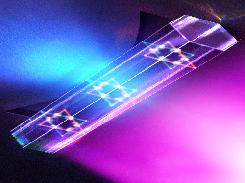 Quantum Laser Turns Energy Loss into Gain
A new laser that generates quantum particles can recycle lost energy for highly efficient, low threshold laser applications
Scientists at KAIST have fabricated a laser system that generates highly interactive quantum particles at room temperature. Their findings, published in the journal Nature Photonics, could lead to a single microcavity laser system that requires lower threshold energy as its energy loss increases.
The system, developed by KAIST physicist Yong-Hoon Cho and colleagues, involves shining light through a single hexagonal-shaped microcavity treated with a loss-modulated silicon nitride substrate. The system design leads to the generation of a polariton laser at room temperature, which is exciting because this usually requires cryogenic temperatures.
The researchers found another unique and counter-intuitive feature of this design. Normally, energy is lost during laser operation. But in this system, as energy loss increased, the amount of energy needed to induce lasing decreased. Exploiting this phenomenon could lead to the development of high efficiency, low threshold lasers for future quantum optical devices.
“This system applies a concept of quantum physics known as parity-time reversal symmetry,” explains Professor Cho. “This is an important platform that allows energy loss to be used as gain. It can be used to reduce laser threshold energy for classical optical devices and sensors, as well as quantum devices and controlling the direction of light.”
The key is the design and materials. The hexagonal microcavity divides light particles into two different modes: one that passes through the upward-facing triangle of the hexagon and another that passes through its downward-facing triangle. Both modes of light particles have the same energy and path but don’t interact with each other.
However, the light particles do interact with other particles called excitons, provided by the hexagonal microcavity, which is made of semiconductors. This interaction leads to the generation of new quantum particles called polaritons that then interact with each other to generate the polariton laser. By controlling the degree of loss between the microcavity and the semiconductor substrate, an intriguing phenomenon arises, with the threshold energy becoming smaller as energy loss increases. This research was supported by the Samsung Science and Technology Foundation and Korea’s National Research Foundation.
-PublicationSong,H.G, Choi, M, Woo, K.Y. Yong-Hoon Cho Room-temperature polaritonic non-Hermitian system with single microcavityNature Photonics (https://doi.org/10.1038/s41566-021-00820-z)
-ProfileProfessor Yong-Hoon ChoQuantum & Nanobio Photonics Laboratoryhttp://qnp.kaist.ac.kr/
Department of PhysicsKAIST
2021.07.07 View 11604
Quantum Laser Turns Energy Loss into Gain
A new laser that generates quantum particles can recycle lost energy for highly efficient, low threshold laser applications
Scientists at KAIST have fabricated a laser system that generates highly interactive quantum particles at room temperature. Their findings, published in the journal Nature Photonics, could lead to a single microcavity laser system that requires lower threshold energy as its energy loss increases.
The system, developed by KAIST physicist Yong-Hoon Cho and colleagues, involves shining light through a single hexagonal-shaped microcavity treated with a loss-modulated silicon nitride substrate. The system design leads to the generation of a polariton laser at room temperature, which is exciting because this usually requires cryogenic temperatures.
The researchers found another unique and counter-intuitive feature of this design. Normally, energy is lost during laser operation. But in this system, as energy loss increased, the amount of energy needed to induce lasing decreased. Exploiting this phenomenon could lead to the development of high efficiency, low threshold lasers for future quantum optical devices.
“This system applies a concept of quantum physics known as parity-time reversal symmetry,” explains Professor Cho. “This is an important platform that allows energy loss to be used as gain. It can be used to reduce laser threshold energy for classical optical devices and sensors, as well as quantum devices and controlling the direction of light.”
The key is the design and materials. The hexagonal microcavity divides light particles into two different modes: one that passes through the upward-facing triangle of the hexagon and another that passes through its downward-facing triangle. Both modes of light particles have the same energy and path but don’t interact with each other.
However, the light particles do interact with other particles called excitons, provided by the hexagonal microcavity, which is made of semiconductors. This interaction leads to the generation of new quantum particles called polaritons that then interact with each other to generate the polariton laser. By controlling the degree of loss between the microcavity and the semiconductor substrate, an intriguing phenomenon arises, with the threshold energy becoming smaller as energy loss increases. This research was supported by the Samsung Science and Technology Foundation and Korea’s National Research Foundation.
-PublicationSong,H.G, Choi, M, Woo, K.Y. Yong-Hoon Cho Room-temperature polaritonic non-Hermitian system with single microcavityNature Photonics (https://doi.org/10.1038/s41566-021-00820-z)
-ProfileProfessor Yong-Hoon ChoQuantum & Nanobio Photonics Laboratoryhttp://qnp.kaist.ac.kr/
Department of PhysicsKAIST
2021.07.07 View 11604 -
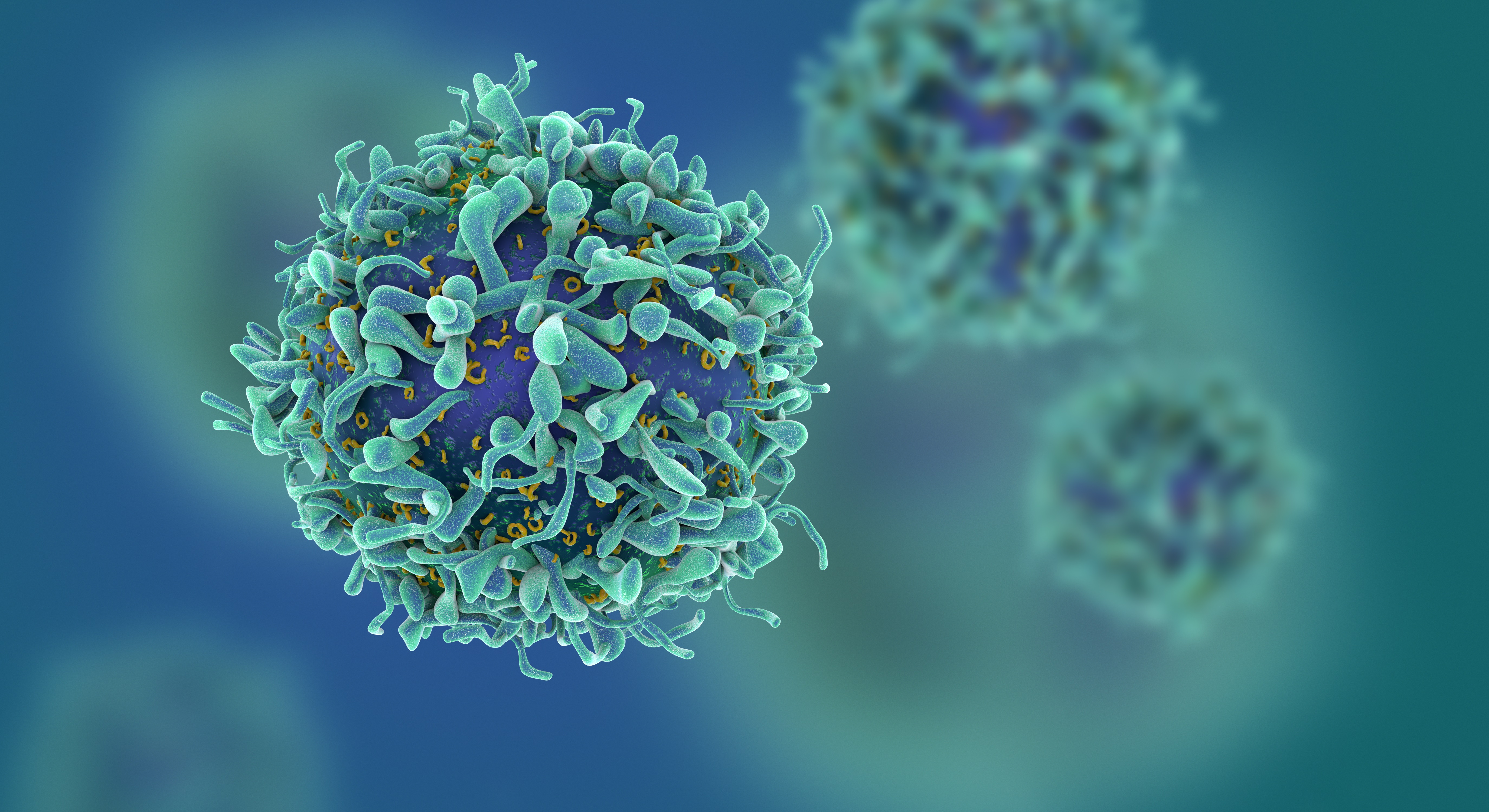 Study of T Cells from COVID-19 Convalescents Guides Vaccine Strategies
Researchers confirm that most COVID-19 patients in their convalescent stage carry stem cell-like memory T cells for months
A KAIST immunology research team found that most convalescent patients of COVID-19 develop and maintain T cell memory for over 10 months regardless of the severity of their symptoms. In addition, memory T cells proliferate rapidly after encountering their cognate antigen and accomplish their multifunctional roles. This study provides new insights for effective vaccine strategies against COVID-19, considering the self-renewal capacity and multipotency of memory T cells.
COVID-19 is a disease caused by severe acute respiratory syndrome coronavirus-2 (SARS-CoV-2) infection. When patients recover from COVID-19, SARS-CoV-2-specific adaptive immune memory is developed. The adaptive immune system consists of two principal components: B cells that produce antibodies and T cells that eliminate infected cells. The current results suggest that the protective immune function of memory T cells will be implemented upon re-exposure to SARS-CoV-2.
Recently, the role of memory T cells against SARS-CoV-2 has been gaining attention as neutralizing antibodies wane after recovery. Although memory T cells cannot prevent the infection itself, they play a central role in preventing the severe progression of COVID-19. However, the longevity and functional maintenance of SARS-CoV-2-specific memory T cells remain unknown.
Professor Eui-Cheol Shin and his collaborators investigated the characteristics and functions of stem cell-like memory T cells, which are expected to play a crucial role in long-term immunity. Researchers analyzed the generation of stem cell-like memory T cells and multi-cytokine producing polyfunctional memory T cells, using cutting-edge immunological techniques.
This research is significant in that revealing the long-term immunity of COVID-19 convalescent patients provides an indicator regarding the long-term persistence of T cell immunity, one of the main goals of future vaccine development, as well as evaluating the long-term efficacy of currently available COVID-19 vaccines.
The research team is presently conducting a follow-up study to identify the memory T cell formation and functional characteristics of those who received COVID-19 vaccines, and to understand the immunological effect of COVID-19 vaccines by comparing the characteristics of memory T cells from vaccinated individuals with those of COVID-19 convalescent patients.
PhD candidate Jae Hyung Jung and Dr. Min-Seok Rha, a clinical fellow at Yonsei Severance Hospital, who led the study together explained, “Our analysis will enhance the understanding of COVID-19 immunity and establish an index for COVID-19 vaccine-induced memory T cells.”
“This study is the world’s longest longitudinal study on differentiation and functions of memory T cells among COVID-19 convalescent patients. The research on the temporal dynamics of immune responses has laid the groundwork for building a strategy for next-generation vaccine development,” Professor Shin added. This work was supported by the Samsung Science and Technology Foundation and KAIST, and was published in Nature Communications on June 30.
-Publication:
Jung, J.H., Rha, MS., Sa, M. et al. SARS-CoV-2-specific T cell memory is sustained in COVID-19 convalescent patients for 10 months with successful development of stem cell-like memory T cells. Nat Communications 12, 4043 (2021). https://doi.org/10.1038/s41467-021-24377-1
-Profile:
Professor Eui-Cheol Shin
Laboratory of Immunology & Infectious Diseases (http://liid.kaist.ac.kr/)
Graduate School of Medical Science and Engineering
KAIST
2021.07.05 View 15007
Study of T Cells from COVID-19 Convalescents Guides Vaccine Strategies
Researchers confirm that most COVID-19 patients in their convalescent stage carry stem cell-like memory T cells for months
A KAIST immunology research team found that most convalescent patients of COVID-19 develop and maintain T cell memory for over 10 months regardless of the severity of their symptoms. In addition, memory T cells proliferate rapidly after encountering their cognate antigen and accomplish their multifunctional roles. This study provides new insights for effective vaccine strategies against COVID-19, considering the self-renewal capacity and multipotency of memory T cells.
COVID-19 is a disease caused by severe acute respiratory syndrome coronavirus-2 (SARS-CoV-2) infection. When patients recover from COVID-19, SARS-CoV-2-specific adaptive immune memory is developed. The adaptive immune system consists of two principal components: B cells that produce antibodies and T cells that eliminate infected cells. The current results suggest that the protective immune function of memory T cells will be implemented upon re-exposure to SARS-CoV-2.
Recently, the role of memory T cells against SARS-CoV-2 has been gaining attention as neutralizing antibodies wane after recovery. Although memory T cells cannot prevent the infection itself, they play a central role in preventing the severe progression of COVID-19. However, the longevity and functional maintenance of SARS-CoV-2-specific memory T cells remain unknown.
Professor Eui-Cheol Shin and his collaborators investigated the characteristics and functions of stem cell-like memory T cells, which are expected to play a crucial role in long-term immunity. Researchers analyzed the generation of stem cell-like memory T cells and multi-cytokine producing polyfunctional memory T cells, using cutting-edge immunological techniques.
This research is significant in that revealing the long-term immunity of COVID-19 convalescent patients provides an indicator regarding the long-term persistence of T cell immunity, one of the main goals of future vaccine development, as well as evaluating the long-term efficacy of currently available COVID-19 vaccines.
The research team is presently conducting a follow-up study to identify the memory T cell formation and functional characteristics of those who received COVID-19 vaccines, and to understand the immunological effect of COVID-19 vaccines by comparing the characteristics of memory T cells from vaccinated individuals with those of COVID-19 convalescent patients.
PhD candidate Jae Hyung Jung and Dr. Min-Seok Rha, a clinical fellow at Yonsei Severance Hospital, who led the study together explained, “Our analysis will enhance the understanding of COVID-19 immunity and establish an index for COVID-19 vaccine-induced memory T cells.”
“This study is the world’s longest longitudinal study on differentiation and functions of memory T cells among COVID-19 convalescent patients. The research on the temporal dynamics of immune responses has laid the groundwork for building a strategy for next-generation vaccine development,” Professor Shin added. This work was supported by the Samsung Science and Technology Foundation and KAIST, and was published in Nature Communications on June 30.
-Publication:
Jung, J.H., Rha, MS., Sa, M. et al. SARS-CoV-2-specific T cell memory is sustained in COVID-19 convalescent patients for 10 months with successful development of stem cell-like memory T cells. Nat Communications 12, 4043 (2021). https://doi.org/10.1038/s41467-021-24377-1
-Profile:
Professor Eui-Cheol Shin
Laboratory of Immunology & Infectious Diseases (http://liid.kaist.ac.kr/)
Graduate School of Medical Science and Engineering
KAIST
2021.07.05 View 15007 -
 Prof. Sang Wan Lee Selected for 2021 IBM Academic Award
Professor Sang Wan Lee from the Department of Bio and Brain Engineering was selected as the recipient of the 2021 IBM Global University Program Academic Award. The award recognizes individual faculty members whose emerging science and technology contains significant interest for universities and IBM.
Professor Lee, whose research focuses on artificial intelligence and computational neuroscience, won the award for his research proposal titled A Neuroscience-Inspired Approach for Metacognitive Reinforcement Learning. IBM provides a gift of $40,000 to the recipient’s institution in recognition of the selection of the project but not as a contract for services.
Professor Lee’s project aims to exploit the unique characteristics of human reinforcement learning. Specifically, he plans to examines the hypothesis that metacognition, a human’s ability to estimate their uncertainty level, serves to guide sample-efficient and near-optimal exploration, making it possible to achieve an optimal balance between model-based and model-free reinforcement learning.
He was also selected as the winner of the Google Research Award in 2016 and has been working with DeepMind and University College London to conduct basic research on decision-making brain science to establish a theory on frontal lobe meta-enhance learning.
"We plan to conduct joint research for utilizing brain-based artificial intelligence technology and frontal lobe meta-enhanced learning technology modeling in collaboration with an international research team including IBM, DeepMind, MIT, and Oxford,” Professor Lee said.
2021.06.25 View 13922
Prof. Sang Wan Lee Selected for 2021 IBM Academic Award
Professor Sang Wan Lee from the Department of Bio and Brain Engineering was selected as the recipient of the 2021 IBM Global University Program Academic Award. The award recognizes individual faculty members whose emerging science and technology contains significant interest for universities and IBM.
Professor Lee, whose research focuses on artificial intelligence and computational neuroscience, won the award for his research proposal titled A Neuroscience-Inspired Approach for Metacognitive Reinforcement Learning. IBM provides a gift of $40,000 to the recipient’s institution in recognition of the selection of the project but not as a contract for services.
Professor Lee’s project aims to exploit the unique characteristics of human reinforcement learning. Specifically, he plans to examines the hypothesis that metacognition, a human’s ability to estimate their uncertainty level, serves to guide sample-efficient and near-optimal exploration, making it possible to achieve an optimal balance between model-based and model-free reinforcement learning.
He was also selected as the winner of the Google Research Award in 2016 and has been working with DeepMind and University College London to conduct basic research on decision-making brain science to establish a theory on frontal lobe meta-enhance learning.
"We plan to conduct joint research for utilizing brain-based artificial intelligence technology and frontal lobe meta-enhanced learning technology modeling in collaboration with an international research team including IBM, DeepMind, MIT, and Oxford,” Professor Lee said.
2021.06.25 View 13922 -
 KAIST-SM Entertainment Joint Research for Metaverse
“Culture scientist will play a role in the future of the entertainment industry”
KAIST President Kwang Hyung Lee and SM Entertainment Founder and Chief Executive Producer Soo-Man Lee signed an MOU on joint research of the metaverse on June 23 at the Daejeon campus. SM Entertainment is the powerhouse of K-pop and Lee is a pioneering figure who helped Korean pop culture emerge into a global phenomenon.
The KAIST-SM metaverse partnership will bring out new culture technology that will lead the virtual entertainment industry by creating more dynamic and vivid digital technologies. KAIST will utilize its AI, robot, and network technologies, while SM will provide its content production expertise for this metaverse research.
President Lee said, “SM artists have mesmerized global audiences and opened new markets for K-Pop. Combining the creativity and cultural imagination of SM with technologies from KAIST, together we will make significant contributions to the advancement of virtual reality as well as the global entertainment industry.”
The Graduate School of Culture Technology has been engaging in a variety of creative projects incorporating science and technology for decades and will now actively participate in this metaverse project with SM.
CEP Lee explained, “The power of celebrities and avatars will rule the future entertainment industry. SM will make a leap forward to be a ‘first mover’ in the digital entertainment industry with this partnership with KAIST. This partnership will shape the new digital future of the entertainment industry boosted by cutting-edge technologies.”
CEP Lee also delivered a special lecture for the KAIST community via Zoom. Saying that producers in the future will be ‘culture scientists’, he stressed the importance of technology converging with culture.
“The key factor for K-pop’s success lies in the impressive technology of Korea. SM places a high priority on developing cultural technology and creating new artists and products combining this technology,” added Lee, citing the hologram contents of Beyond Live concerts and the new 4+4 girl group composed of four girls and four avatars called ‘Aespa.’
2021.06.25 View 8270
KAIST-SM Entertainment Joint Research for Metaverse
“Culture scientist will play a role in the future of the entertainment industry”
KAIST President Kwang Hyung Lee and SM Entertainment Founder and Chief Executive Producer Soo-Man Lee signed an MOU on joint research of the metaverse on June 23 at the Daejeon campus. SM Entertainment is the powerhouse of K-pop and Lee is a pioneering figure who helped Korean pop culture emerge into a global phenomenon.
The KAIST-SM metaverse partnership will bring out new culture technology that will lead the virtual entertainment industry by creating more dynamic and vivid digital technologies. KAIST will utilize its AI, robot, and network technologies, while SM will provide its content production expertise for this metaverse research.
President Lee said, “SM artists have mesmerized global audiences and opened new markets for K-Pop. Combining the creativity and cultural imagination of SM with technologies from KAIST, together we will make significant contributions to the advancement of virtual reality as well as the global entertainment industry.”
The Graduate School of Culture Technology has been engaging in a variety of creative projects incorporating science and technology for decades and will now actively participate in this metaverse project with SM.
CEP Lee explained, “The power of celebrities and avatars will rule the future entertainment industry. SM will make a leap forward to be a ‘first mover’ in the digital entertainment industry with this partnership with KAIST. This partnership will shape the new digital future of the entertainment industry boosted by cutting-edge technologies.”
CEP Lee also delivered a special lecture for the KAIST community via Zoom. Saying that producers in the future will be ‘culture scientists’, he stressed the importance of technology converging with culture.
“The key factor for K-pop’s success lies in the impressive technology of Korea. SM places a high priority on developing cultural technology and creating new artists and products combining this technology,” added Lee, citing the hologram contents of Beyond Live concerts and the new 4+4 girl group composed of four girls and four avatars called ‘Aespa.’
2021.06.25 View 8270 -
 ‘Urban Green Space Affects Citizens’ Happiness’
Study finds the relationship between green space, the economy, and happiness
A recent study revealed that as a city becomes more economically developed, its citizens’ happiness becomes more directly related to the area of urban green space.
A joint research project by Professor Meeyoung Cha of the School of Computing and her collaborators studied the relationship between green space and citizen happiness by analyzing big data from satellite images of 60 different countries.
Urban green space, including parks, gardens, and riversides not only provides aesthetic pleasure, but also positively affects our health by promoting physical activity and social interactions. Most of the previous research attempting to verify the correlation between urban green space and citizen happiness was based on few developed countries. Therefore, it was difficult to identify whether the positive effects of green space are global, or merely phenomena that depended on the economic state of the country. There have also been limitations in data collection, as it is difficult to visit each location or carry out investigations on a large scale based on aerial photographs.
The research team used data collected by Sentinel-2, a high-resolution satellite operated by the European Space Agency (ESA) to investigate 90 green spaces from 60 different countries around the world. The subjects of analysis were cities with the highest population densities (cities that contain at least 10% of the national population), and the images were obtained during the summer of each region for clarity. Images from the northern hemisphere were obtained between June and September of 2018, and those from the southern hemisphere were obtained between December of 2017 and February of 2018.
The areas of urban green space were then quantified and crossed with data from the World Happiness Report and GDP by country reported by the United Nations in 2018. Using these data, the relationships between green space, the economy, and citizen happiness were analyzed.
The results showed that in all cities, citizen happiness was positively correlated with the area of urban green space regardless of the country’s economic state. However, out of the 60 countries studied, the happiness index of the bottom 30 by GDP showed a stronger correlation with economic growth. In countries whose gross national income (GDP per capita) was higher than 38,000 USD, the area of green space acted as a more important factor affecting happiness than economic growth. Data from Seoul was analyzed to represent South Korea, and showed an increased happiness index with increased green areas compared to the past.
The authors point out their work has several policy-level implications. First, public green space should be made accessible to urban dwellers to enhance social support. If public safety in urban parks is not guaranteed, its positive role in social support and happiness may diminish. Also, the meaning of public safety may change; for example, ensuring biological safety will be a priority in keeping urban parks accessible during the COVID-19 pandemic.
Second, urban planning for public green space is needed for both developed and developing countries. As it is challenging or nearly impossible to secure land for green space after the area is developed, urban planning for parks and green space should be considered in developing economies where new cities and suburban areas are rapidly expanding.
Third, recent climate changes can present substantial difficulty in sustaining urban green space. Extreme events such as wildfires, floods, droughts, and cold waves could endanger urban forests while global warming could conversely accelerate tree growth in cities due to the urban heat island effect. Thus, more attention must be paid to predict climate changes and discovering their impact on the maintenance of urban green space.
“There has recently been an increase in the number of studies using big data from satellite images to solve social conundrums,” said Professor Cha. “The tool developed for this investigation can also be used to quantify the area of aquatic environments like lakes and the seaside, and it will now be possible to analyze the relationship between citizen happiness and aquatic environments in future studies,” she added.
Professor Woo Sung Jung from POSTECH and Professor Donghee Wohn from the New Jersey Institute of Technology also joined this research. It was reported in the online issue of EPJ Data Science on May 30.
-PublicationOh-Hyun Kwon, Inho Hong, Jeasurk Yang, Donghee Y. Wohn, Woo-Sung Jung, andMeeyoung Cha, 2021. Urban green space and happiness in developed countries. EPJ Data Science. DOI: https://doi.org/10.1140/epjds/s13688-021-00278-7
-ProfileProfessor Meeyoung ChaData Science Labhttps://ds.ibs.re.kr/
School of Computing
KAIST
2021.06.21 View 13362
‘Urban Green Space Affects Citizens’ Happiness’
Study finds the relationship between green space, the economy, and happiness
A recent study revealed that as a city becomes more economically developed, its citizens’ happiness becomes more directly related to the area of urban green space.
A joint research project by Professor Meeyoung Cha of the School of Computing and her collaborators studied the relationship between green space and citizen happiness by analyzing big data from satellite images of 60 different countries.
Urban green space, including parks, gardens, and riversides not only provides aesthetic pleasure, but also positively affects our health by promoting physical activity and social interactions. Most of the previous research attempting to verify the correlation between urban green space and citizen happiness was based on few developed countries. Therefore, it was difficult to identify whether the positive effects of green space are global, or merely phenomena that depended on the economic state of the country. There have also been limitations in data collection, as it is difficult to visit each location or carry out investigations on a large scale based on aerial photographs.
The research team used data collected by Sentinel-2, a high-resolution satellite operated by the European Space Agency (ESA) to investigate 90 green spaces from 60 different countries around the world. The subjects of analysis were cities with the highest population densities (cities that contain at least 10% of the national population), and the images were obtained during the summer of each region for clarity. Images from the northern hemisphere were obtained between June and September of 2018, and those from the southern hemisphere were obtained between December of 2017 and February of 2018.
The areas of urban green space were then quantified and crossed with data from the World Happiness Report and GDP by country reported by the United Nations in 2018. Using these data, the relationships between green space, the economy, and citizen happiness were analyzed.
The results showed that in all cities, citizen happiness was positively correlated with the area of urban green space regardless of the country’s economic state. However, out of the 60 countries studied, the happiness index of the bottom 30 by GDP showed a stronger correlation with economic growth. In countries whose gross national income (GDP per capita) was higher than 38,000 USD, the area of green space acted as a more important factor affecting happiness than economic growth. Data from Seoul was analyzed to represent South Korea, and showed an increased happiness index with increased green areas compared to the past.
The authors point out their work has several policy-level implications. First, public green space should be made accessible to urban dwellers to enhance social support. If public safety in urban parks is not guaranteed, its positive role in social support and happiness may diminish. Also, the meaning of public safety may change; for example, ensuring biological safety will be a priority in keeping urban parks accessible during the COVID-19 pandemic.
Second, urban planning for public green space is needed for both developed and developing countries. As it is challenging or nearly impossible to secure land for green space after the area is developed, urban planning for parks and green space should be considered in developing economies where new cities and suburban areas are rapidly expanding.
Third, recent climate changes can present substantial difficulty in sustaining urban green space. Extreme events such as wildfires, floods, droughts, and cold waves could endanger urban forests while global warming could conversely accelerate tree growth in cities due to the urban heat island effect. Thus, more attention must be paid to predict climate changes and discovering their impact on the maintenance of urban green space.
“There has recently been an increase in the number of studies using big data from satellite images to solve social conundrums,” said Professor Cha. “The tool developed for this investigation can also be used to quantify the area of aquatic environments like lakes and the seaside, and it will now be possible to analyze the relationship between citizen happiness and aquatic environments in future studies,” she added.
Professor Woo Sung Jung from POSTECH and Professor Donghee Wohn from the New Jersey Institute of Technology also joined this research. It was reported in the online issue of EPJ Data Science on May 30.
-PublicationOh-Hyun Kwon, Inho Hong, Jeasurk Yang, Donghee Y. Wohn, Woo-Sung Jung, andMeeyoung Cha, 2021. Urban green space and happiness in developed countries. EPJ Data Science. DOI: https://doi.org/10.1140/epjds/s13688-021-00278-7
-ProfileProfessor Meeyoung ChaData Science Labhttps://ds.ibs.re.kr/
School of Computing
KAIST
2021.06.21 View 13362