Optical+Society
-
 Microscopy Approach Poised to Offer New Insights into Liver Diseases
Researchers have developed a new way to visualize the progression of nonalcoholic fatty liver disease (NAFLD) in mouse models of the disease. The new microscopy method provides a high-resolution 3D view that could lead to important new insights into NAFLD, a condition in which too much fat is stored in the liver.
“It is estimated that a quarter of the adult global population has NAFLD, yet an effective treatment strategy has not been found,” said professor Pilhan Kim from the Graduate School of Medical Science and Engineering at KAIST. “NAFLD is associated with obesity and type 2 diabetes and can sometimes progress to liver failure in serious case.”
In the Optical Society (OSA) journal Biomedical Optics Express, Professor Kim and colleagues reported their new imaging technique and showed that it can be used to observe how tiny droplets of fat, or lipids, accumulate in the liver cells of living mice over time.
“It has been challenging to find a treatment strategy for NAFLD because most studies examine excised liver tissue that represents just one timepoint in disease progression,” said Professor Kim. “Our technique can capture details of lipid accumulation over time, providing a highly useful research tool for identifying the multiple parameters that likely contribute to the disease and could be targeted with treatment.”
Capturing the dynamics of NAFLD in living mouse models of the disease requires the ability to observe quickly changing interactions of biological components in intact tissue in real-time. To accomplish this, the researchers developed a custom intravital confocal and two-photon microscopy system that acquires images of multiple fluorescent labels at video-rate with cellular resolution.
“With video-rate imaging capability, the continuous movement of liver tissue in live mice due to breathing and heart beating could be tracked in real time and precisely compensated,” said Professor Kim. “This provided motion-artifact free high-resolution images of cellular and sub-cellular sized individual lipid droplets.”
The key to fast imaging was a polygonal mirror that rotated at more than 240 miles per hour to provide extremely fast laser scanning. The researchers also incorporated four different lasers and four high-sensitivity optical detectors into the setup so that they could acquire multi-color images to capture different color fluorescent probes used to label the lipid droplets and microvasculature in the livers of live mice.
“Our approach can capture real-time changes in cell behavior and morphology, vascular structure and function, and the spatiotemporal localization of biological components while directly visualizing of lipid droplet development in NAFLD progression,” said Professor Kim. “It also allows the analysis of the highly complex behaviors of various immune cells as NAFLD progresses.”
The researchers demonstrated their approach by using it to observe the development and spatial distribution of lipid droplets in individual mice with NAFLD induced by a methionine and choline-deficient diet. Next, they plan to use it to study how the liver microenvironment changes during NAFLD progression by imaging the same mouse over time. They also want to use their microscope technique to visualize various immune cells and lipid droplets to better understand the complex liver microenvironment in NAFLD progression.
2020.08.21 View 10604
 -
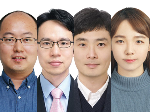
 ‘OSK Rising Stars 30’ Recognizes Four KAISTians
Four KAISTians were selected as star researchers to brighten the future of optics in commemoration of the 30th anniversary of the Optical Society of Korea (OSK). As ‘OSK Rising Stars 30’, the OSK named 27 domestic researchers under the age of 40 who have made significant contributions and will continue contributing to the development of Korea’s optics academia and industry.
Professor YongKeun Park from the Department of Physics was selected in recognition of his contributions to the field of biomedical optics. Professor Park focuses on developing novel optical methods for understanding, diagnosing, and treating human diseases, based on light scattering, light manipulation, and interferometry. As a member of numerous international optics societies including the OSA and the SPIE and a co-founder of two start-up companies, Professor Park continues to broaden his boundaries as a leading opticist and entrepreneur.
Professor Jonghwa Shin from the Department of Materials Science and Engineering was recognized for blazing a trail in the field of broadband metamaterials. Professor Shin’s research on the broadband enhancement of the electric permittivity and refractive index of metamaterials has great potential in both academia and industry.
Professor Hongki Yoo from the Department of Mechanical Engineering is expected to create a significant ripple effect in the diagnosis of cardiovascular disorders through the development of new optical imaging techniques and applications.
Finally, Dr. Sejeong Kim, a KAIST graduate and a Chancellor’s postdoctoral research fellow at the University of Technology Sydney (UTS), was acknowledged for her optical device research utilizing two-dimensional materials. Dr. Kim’s research at UTS now focuses on the introduction of micro/nano cavities for new materials.
(END)
2020.03.16 View 12651
 -
 KAIST Develops Core Technology for Ultra-small 3D Image Sensor
(from left: Dr. Jong-Bum Yo, PhD candidate Seong-Hwan Kimand Professor Hyo-Hoon Park)
A KAIST research team developed a silicon optical phased array (OPA) chip, which can be a core component for three-dimensional image sensors. This research was co-led by PhD candidate Seong-Hwan Kim and Dr. Jong-Bum You from the National Nanofab Center (NNFC).
A 3D image sensor adds distance information to a two-dimensional image, such as a photo, to recognize it as a 3D image. It plays a vital role in various electronics including autonomous vehicles, drones, robots, and facial recognition systems, which require accurate measurement of the distance from objects.
Many automobile and drone companies are focusing on developing 3D image sensor systems, based on mechanical light detection and ranging (LiDAR) systems. However, it can only get as small as the size of a fist and has a high possibility of malfunctioning because it employs a mechanical method for laser beam-steering.
OPAs have gained a great attention as a key component to implement solid-state LiDAR because it can control the light direction electronically without moving parts. Silicon-based OPAs are small, durable, and can be mass-produced through conventional Si-CMOS processes.
However, in the development of OPAs, a big issue has been raised about how to achieve wide beam-steering in transversal and longitudinal directions. In the transversal direction, a wide beam-steering has been implemented, relatively easily, through a thermo-optic or electro-optic control of the phase shifters integrated with a 1D array. But the longitudinal beam-steering has been remaining as a technical challenge since only a narrow steering was possible with the same 1D array by changing the wavelengths of light, which is hard to implement in semiconductor processes.
If a light wavelength is changed, characteristics of element devices consisting the OPA can vary, which makes it difficult to control the light direction with reliability as well as to integrate a wavelength-tunable laser on a silicon-based chip. Therefore, it is essential to devise a new structure that can easily adjust the radiated light in both transversal and longitudinal directions.
By integrating tunable radiator, instead of tunable laser in a conventional OPA, Professor Hyo-Hoon Park from the School of Electrical Engineering and his team developed an ultra-small, low-power OPA chip that facilitates a wide 2D beam-steering with a monochromatic light source.
This OPA structure allows the minimizing of the 3D image sensors, as small as a dragonfly’s eye.
According to the team, the OPA can function as a 3D image sensor and also as a wireless transmitter sending the image data to a desired direction, enabling high-quality image data to be freely communicated between electronic devices.
Kim said, “It’s not an easy task to integrate a tunable light source in the OPA structures of previous works. We hope our research proposing a tunable radiator makes a big step towards commercializing OPAs.”
Dr. You added, “We will be able to support application researches of 3D image sensors, especially for facial recognition with smartphones and augmented reality services. We will try to prepare a processing platform in NNFC that provides core technologies of the 3D image sensor fabrication.”
This research was published in Optics Letters on January 15.
Figure 1.The manufactured OPA chip
Figure 2. Schematic feature showing an application of the OPA to a 3D image sensor
2019.02.08 View 7691
 -
 Professor Jungwon Kim Wins Haerim Optics and Photonics Award
(Professor Jungwon Kim)
Professor Jungwon Kim from the Department of Mechanical Engineering received the 8th Haerim Optics and Photonics Award from the Optical Society of Korea (OSK).
He was recognized for his dedication to pioneering the field of microwave photonics by developing ultra-low noise fiber photonics lasers.
The Haerim Optics and Photonics Award is given to an outstanding researcher who has made academic contributions in the field of optics and photonics for the last five years.
The name of the award (Haerim) comes from the pen-name of the renowned scholar, Professor Un-Chul Paek, because it is maintained using funds he contributed to the OSK.
The OSK will confer the award on February 8 during the 29th OSK Annual Meeting and Winter Conference of 2018.
2018.02.07 View 9009
 -
 Meet the KAISTian of 2017, Professor YongKeun Park
Professor YongKeun Park from the Department of Physics is one of the star professors in KAIST. Rising to the academic stardom, Professor Park’s daily schedule is filled with series of business meetings in addition to lab meetings and lectures.
The year 2017 must have been special for him. During the year, he published numerous papers in international journals, such as Nature Photonics, Nature Communications and Science Advances. These high performances drew international attention from renowned media, including Newsweek and Forbes. Moreover, recognizing his research performance, he was elected as a fellow member of the Optical Society (OSA) in his mid-30s. Noting that the members’ age ranges from late 50s to early 60s, Professor Park’s case considered to be quite exceptional.
Adding to his academic achievement, he has launched two startups powered of his own technologies. One is called Tomocube, a company specialized in 3-D imaging microscope using holotomography technology. His company is currently exporting the products to multiple countries, including the United States and Japan. The other one is The.Wave.Talk which has technologies for examining pre-existing bacteria anywhere and anytime.
His research career and entrepreneurship are well deserved recipient of many honors. At the 2018 kick-off ceremony, Professor Park was awarded the KAISTian of 2017 in recognition of his developing holographic measure and control technology as well as founding a new field for technology application.
KAISTian of the Year, first presented in 2001, is an award to recognize the achievements and exemplary contribution of KAIST member who has put significant effort nationally and internationally, enhancing the value of KAIST.
While receiving the award, he thanked his colleagues and his students who have achieved this far together. He said, “I would like to thank KAIST for providing environment for young professors like me so that we can engage themselves in research. Also, I would like to mention that I am an idea seeder and my students do the most of the research. So, I appreciate my students for their hard works, and it is very pleasure to have them. Lastly, I thank the professors for teaching these outstanding students. I feel great responsibility over this title. I will dedicate myself to make further progress in commercializing technology in KAIST.”
Expecting his successful startup cases as a model and great inspiration to students as well as professors, KAIST interviewed Professor Park.
Q What made you decide to found your startups?
A I believed that my research areas could be further used. As a professor, I believe that it is a university’s role to create added value through commercializing technology and creating startups.
Q You have co-founded two startups. What is your role in each company?
A So, basically I have two full-time jobs, professor in KAIST and CTO in Tomocube. After transferring the technology, I hold the position of advisor in The.Wave.Talk.
(Holographic images captured by the product Professor Park developed)
Q Do your students also participate in your companies or can they?
A No, the school and companies are separate spaces; in other words, they are not participating in my companies. They have trained my employees when transferring the technologies, but they are not directly working for the companies.
However, they can participate if they want to. If there’s a need to develop a certain technology, an industry-academia contract can be made. According to the agreement, students can work for the companies.
Q Were there any hardships when preparing the startups?
A At the initial stage, I did not have a financial problem, thanks to support from Startup KAIST. Yet, inviting capital is the beginning, and I think every step I made to operate, generate revenue, and so on is not easy.
Q Do you believe KAIST is startup-friendly?
A Yes, there’s no school like KAIST in Korea and any other country. Besides various programs to support startup activities, Startup KAIST has many professors equipped with a great deal of experience. Therefore, I believe that KAIST provides an excellent environment for both students and professors to create startups.
Q Do you have any suggestion to KAIST institutionally?
A Well, I would like to make a comment to students and professors in KAIST. I strongly recommend them to challenge themselves by launching startups if they have good ideas. Many students wish to begin their jobs in government-funded research institutes or major corporates, but I believe that engaging in a startup company will also give them valuable and very productive experience.
Unlike before, startup institutions are well established, so attracting good capital is not so hard. There are various activities offered by Startup KAIST, so it’s worthwhile giving it a try.
Q What is your goal for 2018 as a professor and entrepreneur?
A I don’t have a grand plan, but I will work harder to produce good students with new topics in KAIST while adding power to my companies to grow bigger.
By Se Yi Kim from the PR Office
2018.01.03 View 13310
 -
 Professor YongKeun Park Elected as a Fellow of the Optical Society
Professor YongKeun Park, from the Department of Physics at KAIST, was elected as a fellow member of the Optical Society (OSA) in Washington, D.C. on September 12. Fellow membership is given to members who have made a significant contribution to the advancement of optics and photonics.
Professor Park was recognized for his research on digital holography and wavefront control technology.
Professor Park has been producing outstanding research outcomes in the field of holographic technology and light scattering control since joining KAIST in 2010. In particular, he developed and commercialized technology for a holographic telescope. He applied it to various medical and biological research projects, leading the field worldwide.
In the past, cells needed to be dyed with fluorescent materials to capture a 3-D image. However, Professor Park’s holotomography (HT) technology can capture 3-D images of living cells and tissues in real time without color dyeing. This technology allows diversified research in the biological and medical field.
Professor Park established a company, Tomocube, Inc. in 2015 to commercialize the technology. In 2016, he received funding from SoftBank Ventures and Hanmi Pharmaceutical. Currently, major institutes, including MIT, the University of Pittsburgh, the German Cancer Research Center, and Seoul National University Hospital are using his equipment.
Recently, Professor Park and his team developed technology based on light scattering measurements. With this technology, they established a company called The Wave Talk and received funding from various organizations, such as NAVER. Its first product is about to be released.
Professor Park said, “I am glad to become a fellow member based on the research outcomes I produced since I was appointed as a professor at KAIST. I would like to thank the excellent researchers as well as the school for its support. I will devote myself to continuously producing novel outcomes in both basic and applied fields.”
Professor Park has published nearly 100 papers in renowned journals including Nature Photonics, Nature Communications, Science Advances, and Physical Review Letters.
2017.10.18 View 13931
 -
 Next-Generation Holographic Microscope for 3D Live Cell Imaging
KAIST researchers have developed a revolutionary bio-medical imaging tool, the HT-1, to view and analyze cells, which is commercially available.
Professor YongKeun Park of the Physics Department at KAIST and his research team have developed a powerful method for 3D imaging of live cells without staining. The researchers announced the launch of their new microscopic tool, the holotomography (HT)-1, to the global marketplace through a Korean start-up that Professor Park co-founded, TomoCube (www.tomocube.com).
Professor Park is a leading researcher in the field of biophotonics and has dedicated much of his research career to working on digital holographic microscopy technology. He collaborated with TomoCube’s R&D team to develop a state-of-the-art, 2D/3D/4D holographic microscope that would allow a real-time label-free visualization of biological cells and tissues.
The HT is an optical analogy of X-ray computed tomography (CT). Both X-ray CT and HT share the same physical principle—the inverse of wave scattering. The difference is that HT uses laser illumination whereas X-ray CT uses X-ray beams. From the measurement of multiple 2D holograms of a cell, coupled with various angles of laser illuminations, the 3D refractive index (RI) distribution of the cell can be reconstructed. The reconstructed 3D RI map provides structural and chemical information of the cell including mass, morphology, protein concentration, and dynamics of the cellular membrane.
The HT enables users to quantitatively and non-invasively investigate the intrinsic properties of biological cells, for example, dry mass and protein concentration. Some of the research team’s breakthroughs that have leveraged HT’s unique and special capabilities can be found in several recent publications, including a lead article on the simultaneous 3D visualization and position tracking of optically trapped particles which was published in Optica on April 20, 2015.
Current fluorescence confocal microscopy techniques require the use of exogenous labeling agents to render high-contrast molecular information. Therefore, drawbacks include possible photo-bleaching, photo-toxicity, and interference with normal molecular activities. Immune or stem cells that need to be reinjected into the body are considered particularly difficult to employ with fluorescence microscopy.
“As one of the two currently available, high-resolution tomographic microscopes in the world, I believe that the HT-1 is the best in class regarding specifications and functionality. Users can see 3D/4D live images of cells, without fixing, coating or staining cells. Sample preparation times are reduced from a few days or hours to just a few minutes,” said Professor Park.
Two Korean hospitals, Seoul National University Hospital in Bundang and Boramae Hospital in Seoul, are using this microscope currently. The research team has also introduced the HT-1 at the Photonics West Exhibition 2016 that took place on February 16-18 in San Francisco, USA.
Professor Park added, “Our technology has set a new paradigm for cell observation under a microscope. I expect that this tomographic microscopy will be more widely used in future in various areas of pharmaceuticals, neuroscience, immunology, hematology, and cell biology.”
Figure 1: HT-1 and Its Specifications
Figure 2: 3D Images of Representative Biological Cells Taken with the HT-1
2016.03.29 View 13939
 -
 Dr. Se-Jung Kim Receives the Grand Prize at the International Photo and Image Contest on Light
Dr. Se-Jung Kim of the Physics Department at KAIST received the Grand Prize at the 2015 Photo and Image Contest of the International Year of Light and Light-based Technologies.
The United Nations has designated the year 2015 as the International Year of Light and Light-based Technologies.
The Optical Society of Korea celebrated the UN’s designation by hosting an international photo and image contest on the theme of light and optics related technology.
Dr. Kim presented a photo of images taken from a liquid crystal, which was entitled “A Micro Pinwheel.” She took pictures of liquid crystal images with a polarizing microscope and then colored the pictures. The liquid crystal has self-assembled circle domain structures, and each domain can form vortex optics. Her adviser for the project is Professor Yong-Hoon Cho of the Physics Department.
Her work was exhibited during the annual conference of the Optical Society of Korea, which was held on July 13-15, 2015 at Gyeong-Ju Hwabaek International Convention Center. It will also be exhibited at the National Science Museum in Gwacheon and the Kim Dae-Jung Convention Center in Gwangju.
Picture: A Micro Pinwheel
2015.07.31 View 11973
 -
 Fast, Accurate 3D Imaging to Track Optically-Trapped Particles
KAIST researchers published an article on the development of a novel technique to precisely track the 3-D positions of optically-trapped particles having complicated geometry in high speed in the April 2015 issue of Optica.
Optical tweezers have been used as an invaluable tool for exerting micro-scale force on microscopic particles and manipulating three-dimensional (3-D) positions of particles. Optical tweezers employ a tightly-focused laser whose beam diameter is smaller than one micrometer (1/100 of hair thickness), which generates attractive force on neighboring microscopic particles moving toward the beam focus. Controlling the positions of the beam focus enabled researchers to hold the particles and move them freely to other locations so they coined the name “optical tweezers.”
To locate the optically-trapped particles by a laser beam, optical microscopes have usually been employed. Optical microscopes measure light signals scattered by the optically-trapped microscopic particles and the positions of the particles in two dimensions. However, it was difficult to quantify the particles’ precise positions along the optic axis, the direction of the beam, from a single image, which is analogous to the difficulty of determining the front and rear positions of objects when closing an eye due to a lack of depth perception. Furthermore, it became more difficult to measure precisely 3-D positions of particles when scattered light signals were distorted by optically-trapped particles having complicated shapes or other particles occlude the target object along the optic axis.
Professor YongKeun Park and his research team in the Department of Physics at the Korea Advanced Institute of Science and Technology (KAIST) employed an optical diffraction tomography (ODT) technique to measure 3-D positions of optically-trapped particles in high speed. The principle of ODT is similar to X-ray CT imaging commonly used in hospitals for visualizing the internal organs of patients. Like X-ray CT imaging, which takes several images from various illumination angles, ODT measures 3-D images of optically-trapped particles by illuminating them with a laser beam in various incidence angles.
The KAIST team used optical tweezers to trap a glass bead with a diameter of 2 micrometers, and moved the bead toward a white blood cell having complicated internal structures. The team measured the 3-D dynamics of the white blood cell as it responded to an approaching glass bead via ODT in the high acquisition rate of 60 images per second. Since the white blood cell screens the glass bead along an optic axis, a conventionally-used optical microscope could not determine the 3-D positions of the glass bead. In contrast, the present method employing ODT localized the 3-D positions of the bead precisely as well as measured the composition of the internal materials of the bead and the white blood cell simultaneously.
Professor Park said, “Our technique has the advantage of measuring the 3-D positions and internal structures of optically-trapped particles in high speed without labelling exogenous fluorescent agents and can be applied in various fields including physics, optics, nanotechnology, and medical science.”
Kyoohyun Kim, the lead author of this paper (“Simultaneous 3D Visualization and Position Tracking of Optically Trapped Particles Using Optical Diffraction Tomography”), added, “This ODT technique can also apply to cellular-level surgeries where optical tweezers are used to manipulate intracellular organelles and to display in real time and in 3-D the images of the reaction of the cell membrane and nucleus during the operation or monitoring the recovery process of the cells from the surgery.”
The research results were published as the cover article in the April 2014 issue of Optica, the newest journal launched last year by the Optical Society of America (OSA) for rapid dissemination of high-impact results related to optics.
Figure 1: This picture shows the concept image of tweezing an optically-trapped glass bead on the cellular membrane of a white blood cell.
Figure 2: High-speed 3-D images produced from optical diffraction tomography technique
2015.04.24 View 12722
 -
 The First Winner of Sang Soo Lee Award in Optics and Photonics
The Optical Society of Korea and the Optical Society of America selected Mario Garavaglia, a researcher at the La Plata Optical Research Center in Argentina, as the first winner of the Sang Soo Lee Award.
Dr. Garavaglia has been selected to receive the award in recognition for his research and education in the field of optics and photonics in Argentina.
The Sang Soo Lee Award, co-established by the Optical Society of Korea and the Optical Society of America in 2012, is awarded to an individual who has made a significant impact in the field. Special considerations are made for individuals who have introduced a new field of research, helped establish a new industry, or made a great contribution to education in the field.
The award is sponsored by the late Doctor Sang Soo Lee's family, the Optical Society of Korea, and the Optical Society of America.
The late Doctor Sang Soo Lee (1925~2010) has been widely known as the 'father of optics' in Korea. He was an active educator, researcher, and writer. Dr. Lee served as the first director of the Korea Advanced Institute of Science (KAIS), the predecessor to KAIST, which was Korea's first research oriented university.
Dr. Lee also served as the 6th president of KAIST between 1989 to 1991 and was a KAIST professor of physics for 21 years. He oversaw the completion of 50 Ph.D. and 100 Master's students as well as published 230 research papers.
Philip Bucksbaum, the president of the Optical Society of America, commented,
"Garavaglia has been an example to the spirit of the Sang Soo Lee Award. The award is the recognition for his tireless efforts and commitment to the development of optics and photonics in Argentina through his teaching, research, and publications."
Jeong-Won Woo, the president of the Optical Society of Korea, said,
"The Sang Soo Lee Award is given to researchers who have consistently contributed to the development of the field. Garavaglia is a well respected researcher in Argentina, and we are truly happy with his selection."
Dr. Garavaglia established a spectroscopy, optic, and laser laboratory in Universidad Nacional de La Plata in 1966. He founded the Center for Optical Research in 1977 and served as the chief of the laboratory until 1991.
Dr. Garavaglia published over 250 research papers in the fields of classical optics, modern optics, photoemission spectroscopy, and laser spectroscopy. He has also received the Galileo Galilei Award from the International Commission for Optics in 1999.
2014.03.31 View 11922
 -
 Seo-Eun Lee, an undergaruate student receives the Best Paper Award from Optical Society of Korea
Seo-Eun Lee, a student studying at KAIST’s Department of Biological Sciences, has won the Best Paper Award from Bio-Photonics Division at the 2014 Optical Society of Korea Winter Conference, held on 19th February at Daejeon Convention Center.
Only one outstanding paper per division is given an award among the total of 270 papers, and it is very unusual for an undergraduate student to win the award in the field that is not her major.
Lee has studied cell imaging using holography technology since June 2013 under the supervision of Professor Yong-Geun Park from the Department of Physics.
The Optical Society of Korea was founded in 1989, and as the largest academy in the field of optics in Korea, it holds academic presentations, seminars and lectures every year.
2014.03.06 View 11229