bio
-
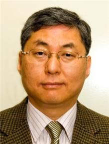
 President Steve Kang of KAIST Attends the 2014 Summer Davos Forum in Tianjin, China
President Steve Kang of KAIST will attend the 2014 Annual Meeting of the New Champions, the World Economic Forum (WEF), to be held on September 10-12, 2014 in Tianjin, China.
KAIST holds its own IdeasLab session on nanotechnology on September 12, 2014.
On September 10, 2014, President Steve Kang will participate in a private session hosted by the Global University Leaders Forum (GULF) community at WEF as a panelist.
In addition to President Kang, eight presidents from top global universities such as the National University of Singapore, Peking University, ETH Zurich (Swiss Federal Institute of Technology), University of Tokyo, and Carnegie Mellon University will join the panel discussion under the topic, “Increasing the Translational Impact of University Research.” Specifically, the presidents will address issues related to the importance of university-led technology transfer in Asia, key strategies and goals for technology transfer, and implementation approaches taken by each university to promote technology transfer from university to industry.
President Kang was invited to this GULF session, the only attendant from Korean universities, in recognition of his long time experience and expertise in education and research.
In 2006, WEF created the GULF, a small community of the presidents of top universities in the world, aiming to offer an open platform for high-level dialogues on issues of higher education and research with other sectors, as well as to foster collaboration between universities in areas of significance for global policy.
As of 2014, a total of 25 globally leading universities, including Harvard University, University of Cambridge, and Massachusetts Institute of Technology, are GULF members. KAIST, which joined the club this year, is the only Korean university.
The 2014 Annual Meeting of the New Champions, also known as the Summer Davos Forum, hosts numerous sessions under the theme of “Creating Value through Innovation.” At the Forum, a total of ten IdeasLab sessions will be hosted. KAIST was invited to run its own IdeasLab on nanotechnology on September 12, 2014.
Together with President Kang, Professors Sang Ouk Kim and Keon Jae Lee from the Department of Materials Science Engineering, KAIST, and Professors Sang Yup Lee and Hyunjoo Lee from the Department of Chemical and Biomolecular Engineering, KAIST, will present their own speeches on the topic entitled “From diagnostics to materials, how is nanotechnology changing lives?”
President Kang will give the opening speech at the KAIST IdeasLab.
He said that an invitation from WEF to join the IdeasLab spoke well for KAIST:
“KAIST is the first and the only Korean university ever invited to run its own IdeasLab at the World Economic Forum. The IdeasLab is an expert group meeting, conducted only by the world’s most prestigious universities and research institutes. At the IdeasLab sessions, global leaders from different sectors identify major issues facing higher education and humanity and explore solutions through science and technology innovation. Holding our own IdeasLab on one of our strongest fields, nanotechnology, is indeed an excellent opportunity for KAIST to show its strength in academic and research excellence on the global stage.”
2014.09.08 View 15882
President Steve Kang of KAIST Attends the 2014 Summer Davos Forum in Tianjin, China
President Steve Kang of KAIST will attend the 2014 Annual Meeting of the New Champions, the World Economic Forum (WEF), to be held on September 10-12, 2014 in Tianjin, China.
KAIST holds its own IdeasLab session on nanotechnology on September 12, 2014.
On September 10, 2014, President Steve Kang will participate in a private session hosted by the Global University Leaders Forum (GULF) community at WEF as a panelist.
In addition to President Kang, eight presidents from top global universities such as the National University of Singapore, Peking University, ETH Zurich (Swiss Federal Institute of Technology), University of Tokyo, and Carnegie Mellon University will join the panel discussion under the topic, “Increasing the Translational Impact of University Research.” Specifically, the presidents will address issues related to the importance of university-led technology transfer in Asia, key strategies and goals for technology transfer, and implementation approaches taken by each university to promote technology transfer from university to industry.
President Kang was invited to this GULF session, the only attendant from Korean universities, in recognition of his long time experience and expertise in education and research.
In 2006, WEF created the GULF, a small community of the presidents of top universities in the world, aiming to offer an open platform for high-level dialogues on issues of higher education and research with other sectors, as well as to foster collaboration between universities in areas of significance for global policy.
As of 2014, a total of 25 globally leading universities, including Harvard University, University of Cambridge, and Massachusetts Institute of Technology, are GULF members. KAIST, which joined the club this year, is the only Korean university.
The 2014 Annual Meeting of the New Champions, also known as the Summer Davos Forum, hosts numerous sessions under the theme of “Creating Value through Innovation.” At the Forum, a total of ten IdeasLab sessions will be hosted. KAIST was invited to run its own IdeasLab on nanotechnology on September 12, 2014.
Together with President Kang, Professors Sang Ouk Kim and Keon Jae Lee from the Department of Materials Science Engineering, KAIST, and Professors Sang Yup Lee and Hyunjoo Lee from the Department of Chemical and Biomolecular Engineering, KAIST, will present their own speeches on the topic entitled “From diagnostics to materials, how is nanotechnology changing lives?”
President Kang will give the opening speech at the KAIST IdeasLab.
He said that an invitation from WEF to join the IdeasLab spoke well for KAIST:
“KAIST is the first and the only Korean university ever invited to run its own IdeasLab at the World Economic Forum. The IdeasLab is an expert group meeting, conducted only by the world’s most prestigious universities and research institutes. At the IdeasLab sessions, global leaders from different sectors identify major issues facing higher education and humanity and explore solutions through science and technology innovation. Holding our own IdeasLab on one of our strongest fields, nanotechnology, is indeed an excellent opportunity for KAIST to show its strength in academic and research excellence on the global stage.”
2014.09.08 View 15882 -
 Kiseok Song, a Ph.D. candidate in the Electrical Engineering Department, receives the 2014 Marconi Society Young Scholar Award
Established
in 1974 to commemorate the eminent Italian inventor and electrical engineer,
Guglielmo Marconi, the Marconi Society has recognized significant contributions
in science and technology by awarding the Marconi Prize, with an annual USD
100,000 grant, to a living scientist who has made great advancements in
communications technology.
Along
with the Marconi Prize, the Society has been presenting the Young Scholars
Awards over the past six years to reward young and emerging scientists’ brilliant
academic and research achievements as well as their entrepreneurship. For this
year’s seventh Young Scholar Awards, a KAIST doctoral student was selected as
one of the two recipients.
Kiseok
Song, a Ph.D. candidate in the Department of Electrical Engineering, KAIST, has
been named as a 2014 Marconi Society Paul Baran Young Scholar. The Marconi Society said that Song was being recognized for "his
academic achievements and leadership in the field of communications and
information science,” according to a press release distributed by the Society on August 28, 2014.
Studying
under the advice of Professor Hoi-Jun Yoo of the Department of Electrical
Engineering at KAIST, Song has developed bio-medical System on a Chip (SoC)
such as smart wireless bio-medical systems combined with optimized SoCs,
compact bio-medical patch systems connected to smart phones, smart electro-acupuncture
and transdermal drug delivery, and multi-modal non-invasive glucose monitors.
The
press release quoted Professor Yoo’s comment on the meaning of Song’s research:
“All
of these bio-medical systems open a new healthcare paradigm to improve people’s
quality of life in combination with the current mobile smart phones.”
In
addition to Song, Himanshu Asnani, a Stanford Ph.D. candidate and system engineer
at Ericsson Silicon Valley, received the other award. The award
ceremony will be held at the Marconi Society’s annual award gala at the
National Academies of Science Building in Washington D.C., on October 2, 2014.
For
details, please read the following press release:
The
Marconi Society, Press Release, August 28, 2014
“Kiseok
Song Receives the 2014 Marconi Society Young Scholar Award”
http://www.marconisociety.org/press/2014Song.html
2014.09.08 View 9924
Kiseok Song, a Ph.D. candidate in the Electrical Engineering Department, receives the 2014 Marconi Society Young Scholar Award
Established
in 1974 to commemorate the eminent Italian inventor and electrical engineer,
Guglielmo Marconi, the Marconi Society has recognized significant contributions
in science and technology by awarding the Marconi Prize, with an annual USD
100,000 grant, to a living scientist who has made great advancements in
communications technology.
Along
with the Marconi Prize, the Society has been presenting the Young Scholars
Awards over the past six years to reward young and emerging scientists’ brilliant
academic and research achievements as well as their entrepreneurship. For this
year’s seventh Young Scholar Awards, a KAIST doctoral student was selected as
one of the two recipients.
Kiseok
Song, a Ph.D. candidate in the Department of Electrical Engineering, KAIST, has
been named as a 2014 Marconi Society Paul Baran Young Scholar. The Marconi Society said that Song was being recognized for "his
academic achievements and leadership in the field of communications and
information science,” according to a press release distributed by the Society on August 28, 2014.
Studying
under the advice of Professor Hoi-Jun Yoo of the Department of Electrical
Engineering at KAIST, Song has developed bio-medical System on a Chip (SoC)
such as smart wireless bio-medical systems combined with optimized SoCs,
compact bio-medical patch systems connected to smart phones, smart electro-acupuncture
and transdermal drug delivery, and multi-modal non-invasive glucose monitors.
The
press release quoted Professor Yoo’s comment on the meaning of Song’s research:
“All
of these bio-medical systems open a new healthcare paradigm to improve people’s
quality of life in combination with the current mobile smart phones.”
In
addition to Song, Himanshu Asnani, a Stanford Ph.D. candidate and system engineer
at Ericsson Silicon Valley, received the other award. The award
ceremony will be held at the Marconi Society’s annual award gala at the
National Academies of Science Building in Washington D.C., on October 2, 2014.
For
details, please read the following press release:
The
Marconi Society, Press Release, August 28, 2014
“Kiseok
Song Receives the 2014 Marconi Society Young Scholar Award”
http://www.marconisociety.org/press/2014Song.html
2014.09.08 View 9924 -
 KAIST Researchers Fabricate Defect-free Graphene for Lithium-ion Batteries
Although graphene has been hailed as promising materials for lithium-ion batteries, making it for large-scale production has remained a challenging task for researchers. So far, high-quality graphene has been produced at the expense of large volume. It is possible to fabricate bulk quantities of graphene, but they will likely contain many defects.
Recently, a KAIST research team, headed by Professors Jung-Ki Park and Hee-Tak Kim from the Department of Chemical and Biomolecular Engineering, developed a fabrication method to produce a large amount of defect-free graphene (df-G) while preserving the structural integrity of the graphene.
This research result was published online in the July 11, 2014 issue of Nano Letters, entitled "Defect-free, Size-tunable Graphene for High-performance Lithium Ion Battery."
Phys.org, a science, research and technology news website, published an article on this research. To read article, please visit the link below:
Phys.org, August 22, 2014
“Scientists fabricate defect-free graphene, set record reversible capacity for Co3O4 node in Li-ion batteries”
http://phys.org/news/2014-08-scientists-fabricate-defect-free-graphene-reversible.html
2014.09.07 View 10682
KAIST Researchers Fabricate Defect-free Graphene for Lithium-ion Batteries
Although graphene has been hailed as promising materials for lithium-ion batteries, making it for large-scale production has remained a challenging task for researchers. So far, high-quality graphene has been produced at the expense of large volume. It is possible to fabricate bulk quantities of graphene, but they will likely contain many defects.
Recently, a KAIST research team, headed by Professors Jung-Ki Park and Hee-Tak Kim from the Department of Chemical and Biomolecular Engineering, developed a fabrication method to produce a large amount of defect-free graphene (df-G) while preserving the structural integrity of the graphene.
This research result was published online in the July 11, 2014 issue of Nano Letters, entitled "Defect-free, Size-tunable Graphene for High-performance Lithium Ion Battery."
Phys.org, a science, research and technology news website, published an article on this research. To read article, please visit the link below:
Phys.org, August 22, 2014
“Scientists fabricate defect-free graphene, set record reversible capacity for Co3O4 node in Li-ion batteries”
http://phys.org/news/2014-08-scientists-fabricate-defect-free-graphene-reversible.html
2014.09.07 View 10682 -
 Extracting Light from Graphite: Core Technology of Graphene Quantum Dots Display Developed
Professor Seokwoo Jeon of the Department of Materials Science and Engineering, Professor Yong-Hoon Cho of the Department of Physics, and Professor Seunghyup Yoo of the Department of Electrical Engineering announced that they were able to develop topnotch graphene quantum dots from graphite.
Using the method of synthesizing graphite intercalation compound from graphite with salt and water, the research team developed graphene quantum dots in an ecofriendly way.
The quantum dots have a diameter of 5 nanometers with their sizes equal and yield high quantum efficiency. Unlike conventional quantum dots, they are not comprised of toxic materials such as lead or cadmium. As the quantum dots can be developed from materials which can be easily found in the nature, researchers look forward to putting these into mass production at low cost.
The research team also discovered a luminescence mechanism of graphene quantum dots and confirmed the possibility of commercial use by developing quantum dot light-emitting diodes with brightness of 1,000 cd/m2, which is greater than that of cellphone displays.
Professor Seokwoo Jeon said, “Although quantum dot LEDs have a lower luminous efficiency than existing ones, their luminescent property can be further improved” and emphasized that “using quantum dot displays will allow us to develop not only paper-thin displays but also flexible ones.”
Sponsored by Graphene Research Center in KAIST Institute for NanoCentury, the research finding was published online in the April 20th issue of Advanced Optical Materials.
Picture 1: Graphene quantum dots and their synthesis
Picture 2: Luminescence mechanism of graphene quantum dots
Picture 3: Structure of graphene quantum dots LED and its emission
2014.09.06 View 19731
Extracting Light from Graphite: Core Technology of Graphene Quantum Dots Display Developed
Professor Seokwoo Jeon of the Department of Materials Science and Engineering, Professor Yong-Hoon Cho of the Department of Physics, and Professor Seunghyup Yoo of the Department of Electrical Engineering announced that they were able to develop topnotch graphene quantum dots from graphite.
Using the method of synthesizing graphite intercalation compound from graphite with salt and water, the research team developed graphene quantum dots in an ecofriendly way.
The quantum dots have a diameter of 5 nanometers with their sizes equal and yield high quantum efficiency. Unlike conventional quantum dots, they are not comprised of toxic materials such as lead or cadmium. As the quantum dots can be developed from materials which can be easily found in the nature, researchers look forward to putting these into mass production at low cost.
The research team also discovered a luminescence mechanism of graphene quantum dots and confirmed the possibility of commercial use by developing quantum dot light-emitting diodes with brightness of 1,000 cd/m2, which is greater than that of cellphone displays.
Professor Seokwoo Jeon said, “Although quantum dot LEDs have a lower luminous efficiency than existing ones, their luminescent property can be further improved” and emphasized that “using quantum dot displays will allow us to develop not only paper-thin displays but also flexible ones.”
Sponsored by Graphene Research Center in KAIST Institute for NanoCentury, the research finding was published online in the April 20th issue of Advanced Optical Materials.
Picture 1: Graphene quantum dots and their synthesis
Picture 2: Luminescence mechanism of graphene quantum dots
Picture 3: Structure of graphene quantum dots LED and its emission
2014.09.06 View 19731 -
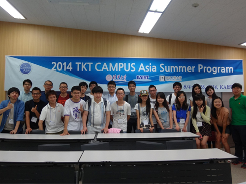 The 2014 CAMPUS Asia Summer Program
The CAMPUS Asia (Collective Action for Mobility Program of University Students in Asia) is an academic exchange program originally proposed at the third presidential meeting of three nations, Korea, China, and Japan, which was held in May 2010 on Jeju Island.
Under the proposal, three science and technology universities, Tsinghua University (China), KAIST (Korea), and Tokyo Institute of Technology (Japan), created a program in 2012 for academic and research collaboration, TKT CAMPUS Asia. Since then, each university has been hosting an exchange program in rotation.
The 2014 summer program of TKT CAMPUS Asia was held from August 4th through 28th at KAIST. A total of 13 professors from the departments of chemical engineering, mechanical engineering, and life science, as well as 25 students participated. Under the summer program, three courses in biotechnology, mechanical engineering, and Korean language were offered. The universities gave credits for all courses.
As part of TKT CAMPUS Asia, KAIST has also operated a semester study abroad and foreign exchange program and will begin a joint degree program by 2015.
Professor Jung Kim of the Department of Mechanical Engineering at KAIST said,
“Just as the Erasmus Program (European Community Action Scheme for the Mobility of University Students) contributed to the foundation of the European Union, we hope that the CAMPUS Asia will serve a similar goal for a greater Asia.”
2014.09.03 View 7213
The 2014 CAMPUS Asia Summer Program
The CAMPUS Asia (Collective Action for Mobility Program of University Students in Asia) is an academic exchange program originally proposed at the third presidential meeting of three nations, Korea, China, and Japan, which was held in May 2010 on Jeju Island.
Under the proposal, three science and technology universities, Tsinghua University (China), KAIST (Korea), and Tokyo Institute of Technology (Japan), created a program in 2012 for academic and research collaboration, TKT CAMPUS Asia. Since then, each university has been hosting an exchange program in rotation.
The 2014 summer program of TKT CAMPUS Asia was held from August 4th through 28th at KAIST. A total of 13 professors from the departments of chemical engineering, mechanical engineering, and life science, as well as 25 students participated. Under the summer program, three courses in biotechnology, mechanical engineering, and Korean language were offered. The universities gave credits for all courses.
As part of TKT CAMPUS Asia, KAIST has also operated a semester study abroad and foreign exchange program and will begin a joint degree program by 2015.
Professor Jung Kim of the Department of Mechanical Engineering at KAIST said,
“Just as the Erasmus Program (European Community Action Scheme for the Mobility of University Students) contributed to the foundation of the European Union, we hope that the CAMPUS Asia will serve a similar goal for a greater Asia.”
2014.09.03 View 7213 -
 KAIST's Advanced Biomass R&D Center and ToolGen will cooperate
The Advanced Biomass R&D Center (ABC) at KAIST and ToolGen, Inc., a Korean biotechnology company focused on the development of engineered nucleases that can be used as essential tools for editing genetic information in microbial, plant, animal, and human cells, signed a memorandum of understanding (MOU) on August 18, 2014 for technology exchange and research collaboration.
ABC is headed by Executive Director Ji-Won Yang, a professor emeritus at the Department of Chemical and Biomolecular Engineering, and Chief Executive Officer Jong-Moon Kim for ToolGen.
The newly signed MOU encourages collaborations in the following areas:
- Development of genome editing technology for microalgae modification
- Development of microalgae that increases biofuel production through the
application of genome editing technology
- Creation of education and training programs for researchers
- Collaboration in other areas
In addition, the two organizations decided to cooperate in the improvement of biofuel yields using ToolGen’s genome editing technology, the commercialization of research outcomes, and the development of eco-friendly biofuels from biomass.
Executive Director Yang commented that “improving biofuel production is crucial to accelerate the commercialization of biofuels, and collaborating with ToolGen will help us realize that goal.” He further said that “The importance of this MOU lies in the fact that the global chemical industry including Korea has been making substantial efforts to shift its attention from a fossil fuel-based development to a more bio-based technology.”
Jin-Soo Kim, the director of the Genome Editing Research Center at the Institute of Basic Sciences in Korea and the cofounder of ToolGen, added that “ToolGen has successfully commercialized its third generation genetic scissors, which shows a lot of promise for commercialization. Our collaboration with KAIST will serve as the driving force to create new industries and accordingly, new jobs.”
2014.09.03 View 11894
KAIST's Advanced Biomass R&D Center and ToolGen will cooperate
The Advanced Biomass R&D Center (ABC) at KAIST and ToolGen, Inc., a Korean biotechnology company focused on the development of engineered nucleases that can be used as essential tools for editing genetic information in microbial, plant, animal, and human cells, signed a memorandum of understanding (MOU) on August 18, 2014 for technology exchange and research collaboration.
ABC is headed by Executive Director Ji-Won Yang, a professor emeritus at the Department of Chemical and Biomolecular Engineering, and Chief Executive Officer Jong-Moon Kim for ToolGen.
The newly signed MOU encourages collaborations in the following areas:
- Development of genome editing technology for microalgae modification
- Development of microalgae that increases biofuel production through the
application of genome editing technology
- Creation of education and training programs for researchers
- Collaboration in other areas
In addition, the two organizations decided to cooperate in the improvement of biofuel yields using ToolGen’s genome editing technology, the commercialization of research outcomes, and the development of eco-friendly biofuels from biomass.
Executive Director Yang commented that “improving biofuel production is crucial to accelerate the commercialization of biofuels, and collaborating with ToolGen will help us realize that goal.” He further said that “The importance of this MOU lies in the fact that the global chemical industry including Korea has been making substantial efforts to shift its attention from a fossil fuel-based development to a more bio-based technology.”
Jin-Soo Kim, the director of the Genome Editing Research Center at the Institute of Basic Sciences in Korea and the cofounder of ToolGen, added that “ToolGen has successfully commercialized its third generation genetic scissors, which shows a lot of promise for commercialization. Our collaboration with KAIST will serve as the driving force to create new industries and accordingly, new jobs.”
2014.09.03 View 11894 -
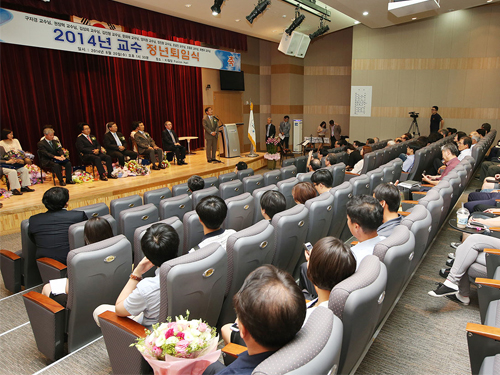 Retiring Faculty Honored
This year’s retirement ceremony took place on August 20, 2014 at Fusion Hall, KI Building, on campus. The ten faculty members whose average service to KAIST exceeded 25 years were honored at the ceremony. They have been appointed emeritus professors this month.
President Steve Kang noted their contributions to the development of KAIST, offering appreciation and recognition of each retiree. The retiring faculty members were:
Ja-Kyung Koo, Professor of Mathematical Sciences
Jang-Hyuk Kwon, Professor of Aerospace Engineering
Sung-Hee Kim, Professor of College of Business
Jin-Hyung Kim, Professor of Computer Science
Hie-Tae Moon, Professor of Physics
Ji-Won Yang, Professor of Chemical and Biomolecular Engineering
Chin-Wan Chung, Professor of Computer Science
Nam-Zin Cho, Professor of Nuclear and Quantum Engineering
Chul-Oh Cho, Professor of Biological Sciences
Byoung-Kyu Choi, Professor of Industrial and Systems Engineering
2014.08.23 View 6858
Retiring Faculty Honored
This year’s retirement ceremony took place on August 20, 2014 at Fusion Hall, KI Building, on campus. The ten faculty members whose average service to KAIST exceeded 25 years were honored at the ceremony. They have been appointed emeritus professors this month.
President Steve Kang noted their contributions to the development of KAIST, offering appreciation and recognition of each retiree. The retiring faculty members were:
Ja-Kyung Koo, Professor of Mathematical Sciences
Jang-Hyuk Kwon, Professor of Aerospace Engineering
Sung-Hee Kim, Professor of College of Business
Jin-Hyung Kim, Professor of Computer Science
Hie-Tae Moon, Professor of Physics
Ji-Won Yang, Professor of Chemical and Biomolecular Engineering
Chin-Wan Chung, Professor of Computer Science
Nam-Zin Cho, Professor of Nuclear and Quantum Engineering
Chul-Oh Cho, Professor of Biological Sciences
Byoung-Kyu Choi, Professor of Industrial and Systems Engineering
2014.08.23 View 6858 -
 ASPIRE League 2014: E-Olympics among Five Asian Universities
About 150 undergraduate students from five leading science and technology (S&T) universities in Asia met at the KAIST campus to attend the E-Olympics on August 7-9, 2014.
The E-Olympics began as a student exchange conference held under the Asian Science and Technology Pioneering Institutes of Research and Education (ASPIRE) League, which offers a variety of events, such as workshops, sports matches, lab visits, special lectures, and art performances, to promote academic and research collaborations and cultural sharing between the students of the league member universities.
Founded in 2009, the ASPIRE League is a university consortium consisted of five top S&T universities in Asia: KAIST in Korea, the Hong Kong University of Science and Technology (HKUST) and Tsinghua University in China, Nanyang Technological University (NTU) in Singapore, and Tokyo Institute of Technology (Tokyo Tech) in Japan. The ASPIRE League aims to provide a knowledge and technology hub for innovation in Asia through the advancement of science and technology and the development of human resources.
Since its start, the ASPIRE League has been holding an annual conference with programs for research collaboration, student exchange, educational cooperation, and satellite laboratories among professors, senior managers, and students of the member universities. This year, however, the consortium decided to dedicate the conference to students by holding the E-Olympics.
Each university sent 30 students to KAIST for the participation of the E-Olympics. For three days, participating students engaged in discussions and presentations at academic workshops; held athletic games including a relay race, basketball, and a rowing race; and toured a few KAIST laboratories, among them: the E-mobility Research Center, the Bio-imaging and Cell Signaling Research Center, the Mechatronics Systems and Control Center, and the Center of Field Robotics for Innovation, Exploration and Defense.
The students also attended a music concert performed by a KAIST student club and a lecture entitled “Entrepreneurship through Global Networking” that emphasized the importance of personnel networking in transferring technological innovation into business opportunities.
Chang-Dong Yoo, the Dean of the International Office at KAIST, said, “The E-Olympics will offer students from top science and technology universities in Asia opportunities to interact with each other on a more personal level. I hope that through many of the E-Olympics programs, the students will learn about each other’s culture and academic strength and develop a sense of community to create a “New Asia” by working together.”
2014.08.11 View 14321
ASPIRE League 2014: E-Olympics among Five Asian Universities
About 150 undergraduate students from five leading science and technology (S&T) universities in Asia met at the KAIST campus to attend the E-Olympics on August 7-9, 2014.
The E-Olympics began as a student exchange conference held under the Asian Science and Technology Pioneering Institutes of Research and Education (ASPIRE) League, which offers a variety of events, such as workshops, sports matches, lab visits, special lectures, and art performances, to promote academic and research collaborations and cultural sharing between the students of the league member universities.
Founded in 2009, the ASPIRE League is a university consortium consisted of five top S&T universities in Asia: KAIST in Korea, the Hong Kong University of Science and Technology (HKUST) and Tsinghua University in China, Nanyang Technological University (NTU) in Singapore, and Tokyo Institute of Technology (Tokyo Tech) in Japan. The ASPIRE League aims to provide a knowledge and technology hub for innovation in Asia through the advancement of science and technology and the development of human resources.
Since its start, the ASPIRE League has been holding an annual conference with programs for research collaboration, student exchange, educational cooperation, and satellite laboratories among professors, senior managers, and students of the member universities. This year, however, the consortium decided to dedicate the conference to students by holding the E-Olympics.
Each university sent 30 students to KAIST for the participation of the E-Olympics. For three days, participating students engaged in discussions and presentations at academic workshops; held athletic games including a relay race, basketball, and a rowing race; and toured a few KAIST laboratories, among them: the E-mobility Research Center, the Bio-imaging and Cell Signaling Research Center, the Mechatronics Systems and Control Center, and the Center of Field Robotics for Innovation, Exploration and Defense.
The students also attended a music concert performed by a KAIST student club and a lecture entitled “Entrepreneurship through Global Networking” that emphasized the importance of personnel networking in transferring technological innovation into business opportunities.
Chang-Dong Yoo, the Dean of the International Office at KAIST, said, “The E-Olympics will offer students from top science and technology universities in Asia opportunities to interact with each other on a more personal level. I hope that through many of the E-Olympics programs, the students will learn about each other’s culture and academic strength and develop a sense of community to create a “New Asia” by working together.”
2014.08.11 View 14321 -
 Newsweek: The Goosebump Sensor That Knows How You Feel
Newsweek covered the introduction of the goosebump sensor invented by Professor Young-Ho Cho of the Department of Bio and Brain Engineering at KAIST in an article dated July 27, 2014.
The article entitled “The Goosebump Sensor That Knows How You Feel” explains how the sensor works and reports on the current research and development trends in emotion-sensing technology.
Professor Cho’s research paper was originally published in the journal Applied Physics Letters on June 24, 2014, titled “A Flexible Skin Piloerection Monitoring Sensor."
Newsweek, July 27, 2014
“The Goosebump Sensor That Knows How You Feel”
http://www.newsweek.com/goosebump-sensor-knows-how-you-feel-260689
2014.07.28 View 9237
Newsweek: The Goosebump Sensor That Knows How You Feel
Newsweek covered the introduction of the goosebump sensor invented by Professor Young-Ho Cho of the Department of Bio and Brain Engineering at KAIST in an article dated July 27, 2014.
The article entitled “The Goosebump Sensor That Knows How You Feel” explains how the sensor works and reports on the current research and development trends in emotion-sensing technology.
Professor Cho’s research paper was originally published in the journal Applied Physics Letters on June 24, 2014, titled “A Flexible Skin Piloerection Monitoring Sensor."
Newsweek, July 27, 2014
“The Goosebump Sensor That Knows How You Feel”
http://www.newsweek.com/goosebump-sensor-knows-how-you-feel-260689
2014.07.28 View 9237 -
 The Journal of Clinical Investigation: Researchers Uncover the Secret Lymphatic Identity of the Schlemm's Canal
The Journal of Clinical Investigation (JCI), a peer-reviewed, top-tier medical journal published by the American Society for Clinical Investigation, carried a commentary entitled “Schlemm’s Canal: More Than Meets the Eye, Lymphatics in Disguise” in the July 25, 2014 issue.
In the commentary, the authors compared a research paper (“Lymphatic regular PROX1 determines Schlemm’s canal integrity and identity”) by Professor Gou-Young Koh of the Graduate School of Medical Science and Engineering at KAIST with research work from the University of Helsinki (article entitled “The Schlemm’s canal is a VEGF-C/VEGFR-3 responsive lymphatic-like vessel”).
The JCI released a press statement dated July 25, 2014 on its commentary. It mentioned that glaucoma, one of the leading causes of blindness worldwide, elevates eye pressure owing to poor drainage of aqueous humor. A specialized structure called “Schlemm’s canal” funnels aqueous humor from the eye back into circulation, which is critical to prevent pressure buildup in the eye. The article discussed the role of Schlemm’s canal in the context of lymphatic vascular characteristics by reviewing two research group’s papers back-to-back.
For the full text of the press release, please visit the link below:
Press Release from the Journal of Clinical Investigation, July 25, 2014
“Researchers uncover the secret lymphatic identity of the Schlemm’s canal”
http://www.eurekalert.org/pub_releases/2014-07/joci-rut072414.php
2014.07.28 View 9202
The Journal of Clinical Investigation: Researchers Uncover the Secret Lymphatic Identity of the Schlemm's Canal
The Journal of Clinical Investigation (JCI), a peer-reviewed, top-tier medical journal published by the American Society for Clinical Investigation, carried a commentary entitled “Schlemm’s Canal: More Than Meets the Eye, Lymphatics in Disguise” in the July 25, 2014 issue.
In the commentary, the authors compared a research paper (“Lymphatic regular PROX1 determines Schlemm’s canal integrity and identity”) by Professor Gou-Young Koh of the Graduate School of Medical Science and Engineering at KAIST with research work from the University of Helsinki (article entitled “The Schlemm’s canal is a VEGF-C/VEGFR-3 responsive lymphatic-like vessel”).
The JCI released a press statement dated July 25, 2014 on its commentary. It mentioned that glaucoma, one of the leading causes of blindness worldwide, elevates eye pressure owing to poor drainage of aqueous humor. A specialized structure called “Schlemm’s canal” funnels aqueous humor from the eye back into circulation, which is critical to prevent pressure buildup in the eye. The article discussed the role of Schlemm’s canal in the context of lymphatic vascular characteristics by reviewing two research group’s papers back-to-back.
For the full text of the press release, please visit the link below:
Press Release from the Journal of Clinical Investigation, July 25, 2014
“Researchers uncover the secret lymphatic identity of the Schlemm’s canal”
http://www.eurekalert.org/pub_releases/2014-07/joci-rut072414.php
2014.07.28 View 9202 -
 Discovery of New Therapeutic Targets for Alzheimer's Disease
A Korean research team headed by Professor Dae-Soo Kim of Biological Sciences at KAIST and Dr. Chang-Jun Lee from the Korea Institute of Science and Technology (KIST) successfully identified that reactive astrocytes, commonly observed in brains affected by Alzheimer’s disease, produce abnormal amounts of inhibitory neurotransmitter gamma-Aminobutyric acid (GABA) in reaction to the enzyme Monoamine oxidase B (Mao-B) and release GABA through the Bestrophin-1 channel to suppress the normal signal transmission of brain nerve cells.
By suppressing the GABA production or release from reactive astrocytes, the research team was able to restore the model mice's memory and learning impairment caused by Alzheimer’s disease. This discovery will allow the development of new drugs to treat Alzheimer’s and other related diseases.
The research result was published in the June 29, 2014 edition of Nature Medicine (Title: GABA from Reactive Astrocytes Impairs Memory in Mouse Models of Alzheimer’s Disease).
For details, please read the article below:
Technology News, July 10, 2014
"Discovery of New Drug Targets for Memory Impairment in Alzheimer’s Disease"
http://technews.tmcnet.com/news/2014/07/10/7917811.htm
2014.07.16 View 10269
Discovery of New Therapeutic Targets for Alzheimer's Disease
A Korean research team headed by Professor Dae-Soo Kim of Biological Sciences at KAIST and Dr. Chang-Jun Lee from the Korea Institute of Science and Technology (KIST) successfully identified that reactive astrocytes, commonly observed in brains affected by Alzheimer’s disease, produce abnormal amounts of inhibitory neurotransmitter gamma-Aminobutyric acid (GABA) in reaction to the enzyme Monoamine oxidase B (Mao-B) and release GABA through the Bestrophin-1 channel to suppress the normal signal transmission of brain nerve cells.
By suppressing the GABA production or release from reactive astrocytes, the research team was able to restore the model mice's memory and learning impairment caused by Alzheimer’s disease. This discovery will allow the development of new drugs to treat Alzheimer’s and other related diseases.
The research result was published in the June 29, 2014 edition of Nature Medicine (Title: GABA from Reactive Astrocytes Impairs Memory in Mouse Models of Alzheimer’s Disease).
For details, please read the article below:
Technology News, July 10, 2014
"Discovery of New Drug Targets for Memory Impairment in Alzheimer’s Disease"
http://technews.tmcnet.com/news/2014/07/10/7917811.htm
2014.07.16 View 10269 -
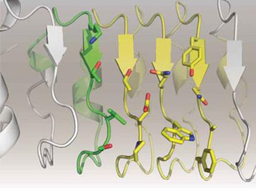 Artificial Antibody-based Therapeutic Candidate for Lung Cancer Developed
Professor Hak-Sung Kim of Biological Sciences at KAIST publishes a cover article on artificial antibody in "Molecular Therapy".
Repebody-based lung cancer therapeutic drug candidate developed Repebody-based protein demonstrates the possibility of the development of a new drug
KAIST Biological Sciences Department’s Professor Hak-Sung Kim, in collaboration with Professor Eun-Kyung Cho from the College of Medicine at Chungnam National University, has successfully developed an artificial antibody-based, or repebody, cancer therapeutic candidate. These research results were published as a cover paper of the July edition of Molecular Therapy.
The repebody developed by Professor Kim and his team strongly binds to interleukin-6, a cancer-causing factor. It has also been confirmed that the repebody can significantly inhibit the proliferation of cancer cells in non-small-cell lung cancer animal model.
Numerous multinational pharmaceutical and biotechnology companies have invested astronomical amounts of money in research for the development of protein therapeutics with low side effects and high efficacy. More than 20 kinds of such therapeutics are currently under clinical trials, and over 100 drugs are under clinical demonstration. Among these, the majority is antibody-based therapeutics, and most of the investments are heavily concentrated in this field. However, antibody production cost is very high because it has large molecular weights and complex structural properties, and this makes it difficult to engineer. Consequently, the development costs a great deal of time and money.
In order to overcome the existing limitations of antibody-based therapeutics, Professor Kim and his team have developed a new artificial antibody, or repebody, which was published in Proceedings of the National Academy of Sciences (PNAS) in 2012. Based on this research, they have succeeded in developing a therapeutic candidate for treating non-small-cell lung cancer with a specifically strong cohesion to the cancer-causing factor, interleukin-6.
Interleukin-6 is a crucial substance within the body that is involved in immune and inflammatory-related signals. When abnormally expressed, it activates various carcinogenic pathways and promotes tumor growth and metastasis. Because of its importance, multinational pharmaceutical companies are heavily investing in developing therapeutics that can inhibit the signaling of interleukin-6.
In this study, Professor Kim and his team observed that a repebody consists of repeated modules, and they conceived a module-based affinity amplification technology that can effectively increase the binding affinity with the disease target. The developed therapeutic candidate has been confirmed in cell and animal experiments to show low immunogenicity, as well as to strongly inhibit the proliferation of non-small-cell lung cancer.
Furthermore, by investigating the complex structure of the repebody with interleukin-6, Professor Kim has identified its mechanism, which demonstrated the potential for therapeutic development. The researchers are currently carrying out pre-clinical trials for acquiring permission to perform clinical trials on animals with non-small-cell lung cancer. The repebody can be developed into a new protein drug after demonstrating its safety and efficacy.
Professor Hak-Sung Kim and his team have confirmed that the repebody can be utilized as a new protein drug, and this will be a significant contribution to Korea’s protein drugs and biotechnology industry development.
The research was supported by the Future Pioneer Industry project and sponsored by the Ministry of Science, ICT and Future Planning.
Figure 1. Professor Kim’s article published as the cover article of July edition of Molecular Therapy
Figure 2. Clinical proof of the repebody’s inhibition of cancer growth using animal models
2014.07.14 View 14332
Artificial Antibody-based Therapeutic Candidate for Lung Cancer Developed
Professor Hak-Sung Kim of Biological Sciences at KAIST publishes a cover article on artificial antibody in "Molecular Therapy".
Repebody-based lung cancer therapeutic drug candidate developed Repebody-based protein demonstrates the possibility of the development of a new drug
KAIST Biological Sciences Department’s Professor Hak-Sung Kim, in collaboration with Professor Eun-Kyung Cho from the College of Medicine at Chungnam National University, has successfully developed an artificial antibody-based, or repebody, cancer therapeutic candidate. These research results were published as a cover paper of the July edition of Molecular Therapy.
The repebody developed by Professor Kim and his team strongly binds to interleukin-6, a cancer-causing factor. It has also been confirmed that the repebody can significantly inhibit the proliferation of cancer cells in non-small-cell lung cancer animal model.
Numerous multinational pharmaceutical and biotechnology companies have invested astronomical amounts of money in research for the development of protein therapeutics with low side effects and high efficacy. More than 20 kinds of such therapeutics are currently under clinical trials, and over 100 drugs are under clinical demonstration. Among these, the majority is antibody-based therapeutics, and most of the investments are heavily concentrated in this field. However, antibody production cost is very high because it has large molecular weights and complex structural properties, and this makes it difficult to engineer. Consequently, the development costs a great deal of time and money.
In order to overcome the existing limitations of antibody-based therapeutics, Professor Kim and his team have developed a new artificial antibody, or repebody, which was published in Proceedings of the National Academy of Sciences (PNAS) in 2012. Based on this research, they have succeeded in developing a therapeutic candidate for treating non-small-cell lung cancer with a specifically strong cohesion to the cancer-causing factor, interleukin-6.
Interleukin-6 is a crucial substance within the body that is involved in immune and inflammatory-related signals. When abnormally expressed, it activates various carcinogenic pathways and promotes tumor growth and metastasis. Because of its importance, multinational pharmaceutical companies are heavily investing in developing therapeutics that can inhibit the signaling of interleukin-6.
In this study, Professor Kim and his team observed that a repebody consists of repeated modules, and they conceived a module-based affinity amplification technology that can effectively increase the binding affinity with the disease target. The developed therapeutic candidate has been confirmed in cell and animal experiments to show low immunogenicity, as well as to strongly inhibit the proliferation of non-small-cell lung cancer.
Furthermore, by investigating the complex structure of the repebody with interleukin-6, Professor Kim has identified its mechanism, which demonstrated the potential for therapeutic development. The researchers are currently carrying out pre-clinical trials for acquiring permission to perform clinical trials on animals with non-small-cell lung cancer. The repebody can be developed into a new protein drug after demonstrating its safety and efficacy.
Professor Hak-Sung Kim and his team have confirmed that the repebody can be utilized as a new protein drug, and this will be a significant contribution to Korea’s protein drugs and biotechnology industry development.
The research was supported by the Future Pioneer Industry project and sponsored by the Ministry of Science, ICT and Future Planning.
Figure 1. Professor Kim’s article published as the cover article of July edition of Molecular Therapy
Figure 2. Clinical proof of the repebody’s inhibition of cancer growth using animal models
2014.07.14 View 14332