TE
-
 Baemin CEO Endows a Scholarship in Honor of the Late Professor Chwa
CEO Beom-Jun Kim of Woowa Brothers also known as ‘Baemin,’ a leading meal delivery app company, made a donation of 100 million KRW in honor of the late Professor Kyong-Yong Chwa from the School of Computing who passed away last year. The fund will be established for the “Kyong-Yong Chwa - Beom-Jun Kim Scholarship” to provide scholarships for four students over five years.
Kim finished his BS in 1997 and MS in 1999 at the School of Computing and Professor Chwa was his advisor. The late Professor Chwa was a pioneering scholar who brought the concept of computer algorithms to Korea. After graduating from Seoul National University in electric engineering, Professor Chwa earned his PhD at Northwestern University and began teaching at KAIST in 1980. Professor Chwa served as the President of the Korean Institute of Information Scientists and Engineers and a fellow emeritus at the Korean Academy of Science and Technology.
Professor Chwa encouraged younger students to participate in international computer programming contests. Under his wing, Team Korea, which was comprised of four high school students, including Kim, placed fourth in the International Olympiad Informatics (IOI). Kim, who participated in the contest as high school junior, won an individual gold medal in the fourth IOI competition in 1992. Since then, Korean students have actively participated in many competitions including the International Collegiate Programming Contest (ICPC) hosted by the Association for Computing Machinery.
Kim said, “I feel fortunate to have met so many good friends and distinguished professors. With them, I had opportunities to grow. I would like to provide such opportunities to my juniors at KAIST. Professor Chwa was a larger than life figure in the field of computer programming. He was always caring and supported us with a warm heart. I want this donation to help carry on his legacy for our students and for them to seek greater challenges and bigger dreams.”
2022.03.25 View 9314
Baemin CEO Endows a Scholarship in Honor of the Late Professor Chwa
CEO Beom-Jun Kim of Woowa Brothers also known as ‘Baemin,’ a leading meal delivery app company, made a donation of 100 million KRW in honor of the late Professor Kyong-Yong Chwa from the School of Computing who passed away last year. The fund will be established for the “Kyong-Yong Chwa - Beom-Jun Kim Scholarship” to provide scholarships for four students over five years.
Kim finished his BS in 1997 and MS in 1999 at the School of Computing and Professor Chwa was his advisor. The late Professor Chwa was a pioneering scholar who brought the concept of computer algorithms to Korea. After graduating from Seoul National University in electric engineering, Professor Chwa earned his PhD at Northwestern University and began teaching at KAIST in 1980. Professor Chwa served as the President of the Korean Institute of Information Scientists and Engineers and a fellow emeritus at the Korean Academy of Science and Technology.
Professor Chwa encouraged younger students to participate in international computer programming contests. Under his wing, Team Korea, which was comprised of four high school students, including Kim, placed fourth in the International Olympiad Informatics (IOI). Kim, who participated in the contest as high school junior, won an individual gold medal in the fourth IOI competition in 1992. Since then, Korean students have actively participated in many competitions including the International Collegiate Programming Contest (ICPC) hosted by the Association for Computing Machinery.
Kim said, “I feel fortunate to have met so many good friends and distinguished professors. With them, I had opportunities to grow. I would like to provide such opportunities to my juniors at KAIST. Professor Chwa was a larger than life figure in the field of computer programming. He was always caring and supported us with a warm heart. I want this donation to help carry on his legacy for our students and for them to seek greater challenges and bigger dreams.”
2022.03.25 View 9314 -
 Tomographic Measurement of Dielectric Tensors
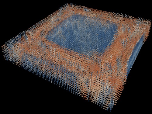
Dielectric tensor tomography allows the direct measurement of the 3D dielectric tensors of optically anisotropic structures
A research team reported the direct measurement of dielectric tensors of anisotropic structures including the spatial variations of principal refractive indices and directors. The group also demonstrated quantitative tomographic measurements of various nematic liquid-crystal structures and their fast 3D nonequilibrium dynamics using a 3D label-free tomographic method. The method was described in Nature Materials.
Light-matter interactions are described by the dielectric tensor. Despite their importance in basic science and applications, it has not been possible to measure 3D dielectric tensors directly. The main challenge was due to the vectorial nature of light scattering from a 3D anisotropic structure. Previous approaches only addressed 3D anisotropic information indirectly and were limited to two-dimensional, qualitative, strict sample conditions or assumptions.
The research team developed a method enabling the tomographic reconstruction of 3D dielectric tensors without any preparation or assumptions. A sample is illuminated with a laser beam with various angles and circularly polarization states. Then, the light fields scattered from a sample are holographically measured and converted into vectorial diffraction components. Finally, by inversely solving a vectorial wave equation, the 3D dielectric tensor is reconstructed.
Professor YongKeun Park said, “There were a greater number of unknowns in direct measuring than with the conventional approach. We applied our approach to measure additional holographic images by slightly tilting the incident angle.”
He said that the slightly tilted illumination provides an additional orthogonal polarization, which makes the underdetermined problem become the determined problem. “Although scattered fields are dependent on the illumination angle, the Fourier differentiation theorem enables the extraction of the same dielectric tensor for the slightly tilted illumination,” Professor Park added.
His team’s method was validated by reconstructing well-known liquid crystal (LC) structures, including the twisted nematic, hybrid aligned nematic, radial, and bipolar configurations. Furthermore, the research team demonstrated the experimental measurements of the non-equilibrium dynamics of annihilating, nucleating, and merging LC droplets, and the LC polymer network with repeating 3D topological defects.
“This is the first experimental measurement of non-equilibrium dynamics and 3D topological defects in LC structures in a label-free manner. Our method enables the exploration of inaccessible nematic structures and interactions in non-equilibrium dynamics,” first author Dr. Seungwoo Shin explained.
-PublicationSeungwoo Shin, Jonghee Eun, Sang Seok Lee, Changjae Lee, Herve Hugonnet, Dong Ki Yoon, Shin-Hyun Kim, Jongwoo Jeong, YongKeun Park, “Tomographic Measurement ofDielectric Tensors at Optical Frequency,” Nature Materials March 02, 2022 (https://doi.org/10/1038/s41563-022-01202-8)
-ProfileProfessor YongKeun ParkBiomedical Optics Laboratory (http://bmol.kaist.ac.kr)Department of PhysicsCollege of Natural SciencesKAIST
2022.03.22 View 9505
Tomographic Measurement of Dielectric Tensors
Dielectric tensor tomography allows the direct measurement of the 3D dielectric tensors of optically anisotropic structures
A research team reported the direct measurement of dielectric tensors of anisotropic structures including the spatial variations of principal refractive indices and directors. The group also demonstrated quantitative tomographic measurements of various nematic liquid-crystal structures and their fast 3D nonequilibrium dynamics using a 3D label-free tomographic method. The method was described in Nature Materials.
Light-matter interactions are described by the dielectric tensor. Despite their importance in basic science and applications, it has not been possible to measure 3D dielectric tensors directly. The main challenge was due to the vectorial nature of light scattering from a 3D anisotropic structure. Previous approaches only addressed 3D anisotropic information indirectly and were limited to two-dimensional, qualitative, strict sample conditions or assumptions.
The research team developed a method enabling the tomographic reconstruction of 3D dielectric tensors without any preparation or assumptions. A sample is illuminated with a laser beam with various angles and circularly polarization states. Then, the light fields scattered from a sample are holographically measured and converted into vectorial diffraction components. Finally, by inversely solving a vectorial wave equation, the 3D dielectric tensor is reconstructed.
Professor YongKeun Park said, “There were a greater number of unknowns in direct measuring than with the conventional approach. We applied our approach to measure additional holographic images by slightly tilting the incident angle.”
He said that the slightly tilted illumination provides an additional orthogonal polarization, which makes the underdetermined problem become the determined problem. “Although scattered fields are dependent on the illumination angle, the Fourier differentiation theorem enables the extraction of the same dielectric tensor for the slightly tilted illumination,” Professor Park added.
His team’s method was validated by reconstructing well-known liquid crystal (LC) structures, including the twisted nematic, hybrid aligned nematic, radial, and bipolar configurations. Furthermore, the research team demonstrated the experimental measurements of the non-equilibrium dynamics of annihilating, nucleating, and merging LC droplets, and the LC polymer network with repeating 3D topological defects.
“This is the first experimental measurement of non-equilibrium dynamics and 3D topological defects in LC structures in a label-free manner. Our method enables the exploration of inaccessible nematic structures and interactions in non-equilibrium dynamics,” first author Dr. Seungwoo Shin explained.
-PublicationSeungwoo Shin, Jonghee Eun, Sang Seok Lee, Changjae Lee, Herve Hugonnet, Dong Ki Yoon, Shin-Hyun Kim, Jongwoo Jeong, YongKeun Park, “Tomographic Measurement ofDielectric Tensors at Optical Frequency,” Nature Materials March 02, 2022 (https://doi.org/10/1038/s41563-022-01202-8)
-ProfileProfessor YongKeun ParkBiomedical Optics Laboratory (http://bmol.kaist.ac.kr)Department of PhysicsCollege of Natural SciencesKAIST
2022.03.22 View 9505 -
 Decoding Brain Signals to Control a Robotic Arm
Advanced brain-machine interface system successfully interprets arm movement directions from neural signals in the brain
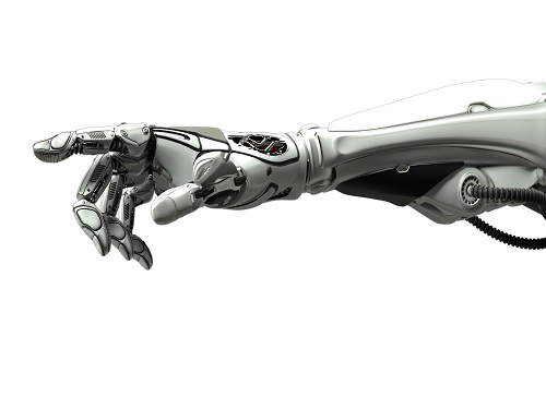
Researchers have developed a mind-reading system for decoding neural signals from the brain during arm movement. The method, described in the journal Applied Soft Computing, can be used by a person to control a robotic arm through a brain-machine interface (BMI).
A BMI is a device that translates nerve signals into commands to control a machine, such as a computer or a robotic limb. There are two main techniques for monitoring neural signals in BMIs: electroencephalography (EEG) and electrocorticography (ECoG).
The EEG exhibits signals from electrodes on the surface of the scalp and is widely employed because it is non-invasive, relatively cheap, safe and easy to use. However, the EEG has low spatial resolution and detects irrelevant neural signals, which makes it difficult to interpret the intentions of individuals from the EEG.
On the other hand, the ECoG is an invasive method that involves placing electrodes directly on the surface of the cerebral cortex below the scalp. Compared with the EEG, the ECoG can monitor neural signals with much higher spatial resolution and less background noise. However, this technique has several drawbacks.
“The ECoG is primarily used to find potential sources of epileptic seizures, meaning the electrodes are placed in different locations for different patients and may not be in the optimal regions of the brain for detecting sensory and movement signals,” explained Professor Jaeseung Jeong, a brain scientist at KAIST. “This inconsistency makes it difficult to decode brain signals to predict movements.”
To overcome these problems, Professor Jeong’s team developed a new method for decoding ECoG neural signals during arm movement. The system is based on a machine-learning system for analysing and predicting neural signals called an ‘echo-state network’ and a mathematical probability model called the Gaussian distribution.
In the study, the researchers recorded ECoG signals from four individuals with epilepsy while they were performing a reach-and-grasp task. Because the ECoG electrodes were placed according to the potential sources of each patient’s epileptic seizures, only 22% to 44% of the electrodes were located in the regions of the brain responsible for controlling movement.
During the movement task, the participants were given visual cues, either by placing a real tennis ball in front of them, or via a virtual reality headset showing a clip of a human arm reaching forward in first-person view. They were asked to reach forward, grasp an object, then return their hand and release the object, while wearing motion sensors on their wrists and fingers. In a second task, they were instructed to imagine reaching forward without moving their arms.
The researchers monitored the signals from the ECoG electrodes during real and imaginary arm movements, and tested whether the new system could predict the direction of this movement from the neural signals. They found that the novel decoder successfully classified arm movements in 24 directions in three-dimensional space, both in the real and virtual tasks, and that the results were at least five times more accurate than chance. They also used a computer simulation to show that the novel ECoG decoder could control the movements of a robotic arm.
Overall, the results suggest that the new machine learning-based BCI system successfully used ECoG signals to interpret the direction of the intended movements. The next steps will be to improve the accuracy and efficiency of the decoder. In the future, it could be used in a real-time BMI device to help people with movement or sensory impairments.
This research was supported by the KAIST Global Singularity Research Program of 2021, Brain Research Program of the National Research Foundation of Korea funded by the Ministry of Science, ICT, and Future Planning, and the Basic Science Research Program through the National Research Foundation of Korea funded by the Ministry of Education.
-PublicationHoon-Hee Kim, Jaeseung Jeong, “An electrocorticographic decoder for arm movement for brain-machine interface using an echo state network and Gaussian readout,” Applied SoftComputing online December 31, 2021 (doi.org/10.1016/j.asoc.2021.108393)
-ProfileProfessor Jaeseung JeongDepartment of Bio and Brain EngineeringCollege of EngineeringKAIST
2022.03.18 View 13477
Decoding Brain Signals to Control a Robotic Arm
Advanced brain-machine interface system successfully interprets arm movement directions from neural signals in the brain
Researchers have developed a mind-reading system for decoding neural signals from the brain during arm movement. The method, described in the journal Applied Soft Computing, can be used by a person to control a robotic arm through a brain-machine interface (BMI).
A BMI is a device that translates nerve signals into commands to control a machine, such as a computer or a robotic limb. There are two main techniques for monitoring neural signals in BMIs: electroencephalography (EEG) and electrocorticography (ECoG).
The EEG exhibits signals from electrodes on the surface of the scalp and is widely employed because it is non-invasive, relatively cheap, safe and easy to use. However, the EEG has low spatial resolution and detects irrelevant neural signals, which makes it difficult to interpret the intentions of individuals from the EEG.
On the other hand, the ECoG is an invasive method that involves placing electrodes directly on the surface of the cerebral cortex below the scalp. Compared with the EEG, the ECoG can monitor neural signals with much higher spatial resolution and less background noise. However, this technique has several drawbacks.
“The ECoG is primarily used to find potential sources of epileptic seizures, meaning the electrodes are placed in different locations for different patients and may not be in the optimal regions of the brain for detecting sensory and movement signals,” explained Professor Jaeseung Jeong, a brain scientist at KAIST. “This inconsistency makes it difficult to decode brain signals to predict movements.”
To overcome these problems, Professor Jeong’s team developed a new method for decoding ECoG neural signals during arm movement. The system is based on a machine-learning system for analysing and predicting neural signals called an ‘echo-state network’ and a mathematical probability model called the Gaussian distribution.
In the study, the researchers recorded ECoG signals from four individuals with epilepsy while they were performing a reach-and-grasp task. Because the ECoG electrodes were placed according to the potential sources of each patient’s epileptic seizures, only 22% to 44% of the electrodes were located in the regions of the brain responsible for controlling movement.
During the movement task, the participants were given visual cues, either by placing a real tennis ball in front of them, or via a virtual reality headset showing a clip of a human arm reaching forward in first-person view. They were asked to reach forward, grasp an object, then return their hand and release the object, while wearing motion sensors on their wrists and fingers. In a second task, they were instructed to imagine reaching forward without moving their arms.
The researchers monitored the signals from the ECoG electrodes during real and imaginary arm movements, and tested whether the new system could predict the direction of this movement from the neural signals. They found that the novel decoder successfully classified arm movements in 24 directions in three-dimensional space, both in the real and virtual tasks, and that the results were at least five times more accurate than chance. They also used a computer simulation to show that the novel ECoG decoder could control the movements of a robotic arm.
Overall, the results suggest that the new machine learning-based BCI system successfully used ECoG signals to interpret the direction of the intended movements. The next steps will be to improve the accuracy and efficiency of the decoder. In the future, it could be used in a real-time BMI device to help people with movement or sensory impairments.
This research was supported by the KAIST Global Singularity Research Program of 2021, Brain Research Program of the National Research Foundation of Korea funded by the Ministry of Science, ICT, and Future Planning, and the Basic Science Research Program through the National Research Foundation of Korea funded by the Ministry of Education.
-PublicationHoon-Hee Kim, Jaeseung Jeong, “An electrocorticographic decoder for arm movement for brain-machine interface using an echo state network and Gaussian readout,” Applied SoftComputing online December 31, 2021 (doi.org/10.1016/j.asoc.2021.108393)
-ProfileProfessor Jaeseung JeongDepartment of Bio and Brain EngineeringCollege of EngineeringKAIST
2022.03.18 View 13477 -
 CXL-Based Memory Disaggregation Technology Opens Up a New Direction for Big Data Solution Frameworks
A KAIST team’s compute express link (CXL) provides new insights on memory disaggregation and ensures direct access and high-performance capabilities
A team from the Computer Architecture and Memory Systems Laboratory (CAMEL) at KAIST presented a new compute express link (CXL) solution whose directly accessible, and high-performance memory disaggregation opens new directions for big data memory processing. Professor Myoungsoo Jung said the team’s technology significantly improves performance compared to existing remote direct memory access (RDMA)-based memory disaggregation.
CXL is a peripheral component interconnect-express (PCIe)-based new dynamic multi-protocol made for efficiently utilizing memory devices and accelerators. Many enterprise data centers and memory vendors are paying attention to it as the next-generation multi-protocol for the era of big data.
Emerging big data applications such as machine learning, graph analytics, and in-memory databases require large memory capacities. However, scaling out the memory capacity via a prior memory interface like double data rate (DDR) is limited by the number of the central processing units (CPUs) and memory controllers. Therefore, memory disaggregation, which allows connecting a host to another host’s memory or memory nodes, has appeared.
RDMA is a way that a host can directly access another host’s memory via InfiniBand, the commonly used network protocol in data centers. Nowadays, most existing memory disaggregation technologies employ RDMA to get a large memory capacity. As a result, a host can share another host’s memory by transferring the data between local and remote memory.
Although RDMA-based memory disaggregation provides a large memory capacity to a host, two critical problems exist. First, scaling out the memory still needs an extra CPU to be added. Since passive memory such as dynamic random-access memory (DRAM), cannot operate by itself, it should be controlled by the CPU. Second, redundant data copies and software fabric interventions for RDMA-based memory disaggregation cause longer access latency. For example, remote memory access latency in RDMA-based memory disaggregation is multiple orders of magnitude longer than local memory access.
To address these issues, Professor Jung’s team developed the CXL-based memory disaggregation framework, including CXL-enabled customized CPUs, CXL devices, CXL switches, and CXL-aware operating system modules. The team’s CXL device is a pure passive and directly accessible memory node that contains multiple DRAM dual inline memory modules (DIMMs) and a CXL memory controller. Since the CXL memory controller supports the memory in the CXL device, a host can utilize the memory node without processor or software intervention. The team’s CXL switch enables scaling out a host’s memory capacity by hierarchically connecting multiple CXL devices to the CXL switch allowing more than hundreds of devices. Atop the switches and devices, the team’s CXL-enabled operating system removes redundant data copy and protocol conversion exhibited by conventional RDMA, which can significantly decrease access latency to the memory nodes.
In a test comparing loading 64B (cacheline) data from memory pooling devices, CXL-based memory disaggregation showed 8.2 times higher data load performance than RDMA-based memory disaggregation and even similar performance to local DRAM memory. In the team’s evaluations for a big data benchmark such as a machine learning-based test, CXL-based memory disaggregation technology also showed a maximum of 3.7 times higher performance than prior RDMA-based memory disaggregation technologies.
“Escaping from the conventional RDMA-based memory disaggregation, our CXL-based memory disaggregation framework can provide high scalability and performance for diverse datacenters and cloud service infrastructures,” said Professor Jung. He went on to stress, “Our CXL-based memory disaggregation research will bring about a new paradigm for memory solutions that will lead the era of big data.”
-Profile: Professor Myoungsoo Jung Computer Architecture and Memory Systems Laboratory (CAMEL)http://camelab.org School of Electrical EngineeringKAIST
2022.03.16 View 24093
CXL-Based Memory Disaggregation Technology Opens Up a New Direction for Big Data Solution Frameworks
A KAIST team’s compute express link (CXL) provides new insights on memory disaggregation and ensures direct access and high-performance capabilities
A team from the Computer Architecture and Memory Systems Laboratory (CAMEL) at KAIST presented a new compute express link (CXL) solution whose directly accessible, and high-performance memory disaggregation opens new directions for big data memory processing. Professor Myoungsoo Jung said the team’s technology significantly improves performance compared to existing remote direct memory access (RDMA)-based memory disaggregation.
CXL is a peripheral component interconnect-express (PCIe)-based new dynamic multi-protocol made for efficiently utilizing memory devices and accelerators. Many enterprise data centers and memory vendors are paying attention to it as the next-generation multi-protocol for the era of big data.
Emerging big data applications such as machine learning, graph analytics, and in-memory databases require large memory capacities. However, scaling out the memory capacity via a prior memory interface like double data rate (DDR) is limited by the number of the central processing units (CPUs) and memory controllers. Therefore, memory disaggregation, which allows connecting a host to another host’s memory or memory nodes, has appeared.
RDMA is a way that a host can directly access another host’s memory via InfiniBand, the commonly used network protocol in data centers. Nowadays, most existing memory disaggregation technologies employ RDMA to get a large memory capacity. As a result, a host can share another host’s memory by transferring the data between local and remote memory.
Although RDMA-based memory disaggregation provides a large memory capacity to a host, two critical problems exist. First, scaling out the memory still needs an extra CPU to be added. Since passive memory such as dynamic random-access memory (DRAM), cannot operate by itself, it should be controlled by the CPU. Second, redundant data copies and software fabric interventions for RDMA-based memory disaggregation cause longer access latency. For example, remote memory access latency in RDMA-based memory disaggregation is multiple orders of magnitude longer than local memory access.
To address these issues, Professor Jung’s team developed the CXL-based memory disaggregation framework, including CXL-enabled customized CPUs, CXL devices, CXL switches, and CXL-aware operating system modules. The team’s CXL device is a pure passive and directly accessible memory node that contains multiple DRAM dual inline memory modules (DIMMs) and a CXL memory controller. Since the CXL memory controller supports the memory in the CXL device, a host can utilize the memory node without processor or software intervention. The team’s CXL switch enables scaling out a host’s memory capacity by hierarchically connecting multiple CXL devices to the CXL switch allowing more than hundreds of devices. Atop the switches and devices, the team’s CXL-enabled operating system removes redundant data copy and protocol conversion exhibited by conventional RDMA, which can significantly decrease access latency to the memory nodes.
In a test comparing loading 64B (cacheline) data from memory pooling devices, CXL-based memory disaggregation showed 8.2 times higher data load performance than RDMA-based memory disaggregation and even similar performance to local DRAM memory. In the team’s evaluations for a big data benchmark such as a machine learning-based test, CXL-based memory disaggregation technology also showed a maximum of 3.7 times higher performance than prior RDMA-based memory disaggregation technologies.
“Escaping from the conventional RDMA-based memory disaggregation, our CXL-based memory disaggregation framework can provide high scalability and performance for diverse datacenters and cloud service infrastructures,” said Professor Jung. He went on to stress, “Our CXL-based memory disaggregation research will bring about a new paradigm for memory solutions that will lead the era of big data.”
-Profile: Professor Myoungsoo Jung Computer Architecture and Memory Systems Laboratory (CAMEL)http://camelab.org School of Electrical EngineeringKAIST
2022.03.16 View 24093 -
 'Fingerprint' Machine Learning Technique Identifies Different Bacteria in Seconds
A synergistic combination of surface-enhanced Raman spectroscopy and deep learning serves as an effective platform for separation-free detection of bacteria in arbitrary media
Bacterial identification can take hours and often longer, precious time when diagnosing infections and selecting appropriate treatments. There may be a quicker, more accurate process according to researchers at KAIST. By teaching a deep learning algorithm to identify the “fingerprint” spectra of the molecular components of various bacteria, the researchers could classify various bacteria in different media with accuracies of up to 98%.
Their results were made available online on Jan. 18 in Biosensors and Bioelectronics, ahead of publication in the journal’s April issue.
Bacteria-induced illnesses, those caused by direct bacterial infection or by exposure to bacterial toxins, can induce painful symptoms and even lead to death, so the rapid detection of bacteria is crucial to prevent the intake of contaminated foods and to diagnose infections from clinical samples, such as urine. “By using surface-enhanced Raman spectroscopy (SERS) analysis boosted with a newly proposed deep learning model, we demonstrated a markedly simple, fast, and effective route to classify the signals of two common bacteria and their resident media without any separation procedures,” said Professor Sungho Jo from the School of Computing.
Raman spectroscopy sends light through a sample to see how it scatters. The results reveal structural information about the sample — the spectral fingerprint — allowing researchers to identify its molecules. The surface-enhanced version places sample cells on noble metal nanostructures that help amplify the sample’s signals.
However, it is challenging to obtain consistent and clear spectra of bacteria due to numerous overlapping peak sources, such as proteins in cell walls. “Moreover, strong signals of surrounding media are also enhanced to overwhelm target signals, requiring time-consuming and tedious bacterial separation steps,” said Professor Yeon Sik Jung from the Department of Materials Science and Engineering.
To parse through the noisy signals, the researchers implemented an artificial intelligence method called deep learning that can hierarchically extract certain features of the spectral information to classify data. They specifically designed their model, named the dual-branch wide-kernel network (DualWKNet), to efficiently learn the correlation between spectral features. Such an ability is critical for analyzing one-dimensional spectral data, according to Professor Jo.
“Despite having interfering signals or noise from the media, which make the general shapes of different bacterial spectra and their residing media signals look similar, high classification accuracies of bacterial types and their media were achieved,” Professor Jo said, explaining that DualWKNet allowed the team to identify key peaks in each class that were almost indiscernible in individual spectra, enhancing the classification accuracies. “Ultimately, with the use of DualWKNet replacing the bacteria and media separation steps, our method dramatically reduces analysis time.”
The researchers plan to use their platform to study more bacteria and media types, using the information to build a training data library of various bacterial types in additional media to reduce the collection and detection times for new samples.
“We developed a meaningful universal platform for rapid bacterial detection with the collaboration between SERS and deep learning,” Professor Jo said. “We hope to extend the use of our deep learning-based SERS analysis platform to detect numerous types of bacteria in additional media that are important for food or clinical analysis, such as blood.”
The National R&D Program, through a National Research Foundation of Korea grant funded by the Ministry of Science and ICT, supported this research.
-PublicationEojin Rho, Minjoon Kim, Seunghee H. Cho, Bongjae Choi, Hyungjoon Park, Hanhwi Jang, Yeon Sik Jung, Sungho Jo, “Separation-free bacterial identification in arbitrary media via deepneural network-based SERS analysis,” Biosensors and Bioelectronics online January 18, 2022 (doi.org/10.1016/j.bios.2022.113991)
-ProfileProfessor Yeon Sik JungDepartment of Materials Science and EngineeringKAIST
Professor Sungho JoSchool of ComputingKAIST
2022.03.04 View 23051
'Fingerprint' Machine Learning Technique Identifies Different Bacteria in Seconds
A synergistic combination of surface-enhanced Raman spectroscopy and deep learning serves as an effective platform for separation-free detection of bacteria in arbitrary media
Bacterial identification can take hours and often longer, precious time when diagnosing infections and selecting appropriate treatments. There may be a quicker, more accurate process according to researchers at KAIST. By teaching a deep learning algorithm to identify the “fingerprint” spectra of the molecular components of various bacteria, the researchers could classify various bacteria in different media with accuracies of up to 98%.
Their results were made available online on Jan. 18 in Biosensors and Bioelectronics, ahead of publication in the journal’s April issue.
Bacteria-induced illnesses, those caused by direct bacterial infection or by exposure to bacterial toxins, can induce painful symptoms and even lead to death, so the rapid detection of bacteria is crucial to prevent the intake of contaminated foods and to diagnose infections from clinical samples, such as urine. “By using surface-enhanced Raman spectroscopy (SERS) analysis boosted with a newly proposed deep learning model, we demonstrated a markedly simple, fast, and effective route to classify the signals of two common bacteria and their resident media without any separation procedures,” said Professor Sungho Jo from the School of Computing.
Raman spectroscopy sends light through a sample to see how it scatters. The results reveal structural information about the sample — the spectral fingerprint — allowing researchers to identify its molecules. The surface-enhanced version places sample cells on noble metal nanostructures that help amplify the sample’s signals.
However, it is challenging to obtain consistent and clear spectra of bacteria due to numerous overlapping peak sources, such as proteins in cell walls. “Moreover, strong signals of surrounding media are also enhanced to overwhelm target signals, requiring time-consuming and tedious bacterial separation steps,” said Professor Yeon Sik Jung from the Department of Materials Science and Engineering.
To parse through the noisy signals, the researchers implemented an artificial intelligence method called deep learning that can hierarchically extract certain features of the spectral information to classify data. They specifically designed their model, named the dual-branch wide-kernel network (DualWKNet), to efficiently learn the correlation between spectral features. Such an ability is critical for analyzing one-dimensional spectral data, according to Professor Jo.
“Despite having interfering signals or noise from the media, which make the general shapes of different bacterial spectra and their residing media signals look similar, high classification accuracies of bacterial types and their media were achieved,” Professor Jo said, explaining that DualWKNet allowed the team to identify key peaks in each class that were almost indiscernible in individual spectra, enhancing the classification accuracies. “Ultimately, with the use of DualWKNet replacing the bacteria and media separation steps, our method dramatically reduces analysis time.”
The researchers plan to use their platform to study more bacteria and media types, using the information to build a training data library of various bacterial types in additional media to reduce the collection and detection times for new samples.
“We developed a meaningful universal platform for rapid bacterial detection with the collaboration between SERS and deep learning,” Professor Jo said. “We hope to extend the use of our deep learning-based SERS analysis platform to detect numerous types of bacteria in additional media that are important for food or clinical analysis, such as blood.”
The National R&D Program, through a National Research Foundation of Korea grant funded by the Ministry of Science and ICT, supported this research.
-PublicationEojin Rho, Minjoon Kim, Seunghee H. Cho, Bongjae Choi, Hyungjoon Park, Hanhwi Jang, Yeon Sik Jung, Sungho Jo, “Separation-free bacterial identification in arbitrary media via deepneural network-based SERS analysis,” Biosensors and Bioelectronics online January 18, 2022 (doi.org/10.1016/j.bios.2022.113991)
-ProfileProfessor Yeon Sik JungDepartment of Materials Science and EngineeringKAIST
Professor Sungho JoSchool of ComputingKAIST
2022.03.04 View 23051 -
 KAA Recognizes 4 Distinguished Alumni of the Year
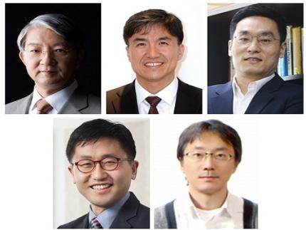
The KAIST Alumni Association (KAA) recognized four distinguished alumni of the year during a ceremony on February 25 in Seoul. The four Distinguished Alumni Awardees are Distinguished Professor Sukbok Chang from the KAIST Department of Chemistry, Hyunshil Ahn, head of the AI Economy Institute and an editorial writer at The Korea Economic Daily, CEO Hwan-ho Sung of PSTech, and President Hark Kyu Park of Samsung Electronics.
Distinguished Professor Sukbok Chang who received his MS from the Department of Chemistry in 1985 has been a pioneer in the novel field of ‘carbon-hydrogen bond activation reactions’. He has significantly contributed to raising Korea’s international reputation in natural sciences and received the Kyungam Academic Award in 2013, the 14th Korea Science Award in 2015, the 1st Science and Technology Prize of Korea Toray in 2018, and the Best Scientist/Engineer Award Korea in 2019. Furthermore, he was named as a Highly Cited Researcher who ranked in the top 1% of citations by field and publication year in the Web of Science citation index for seven consecutive years from 2015 to 2021, demonstrating his leadership as a global scholar.
Hyunshil Ahn, a graduate of the School of Business and Technology Management with an MS in 1985 and a PhD in 1987, was appointed as the first head of the AI Economy Institute when The Korea Economic Daily was the first Korean media outlet to establish an AI economy lab. He has contributed to creating new roles for the press and media in the 4th industrial revolution, and added to the popularization of AI technology through regulation reform and consulting on industrial policies.
PSTech CEO Hwan-ho Sung is a graduate of the School of Electrical Engineering where he received an MS in 1988 and a PhD in EMBA in 2008. He has run the electronics company PSTech for over 20 years and successfully localized the production of power equipment, which previously depended on foreign technology. His development of the world’s first power equipment that can be applied to new industries including semiconductors and displays was recognized through this award.
Samsung Electronics President Hark Kyu Park graduated from the School of Business and Technology Management with an MS in 1986. He not only enhanced Korea’s national competitiveness by expanding the semiconductor industry, but also established contract-based semiconductor departments at Korean universities including KAIST, Sungkyunkwan University, Yonsei University, and Postech, and semiconductor track courses at KAIST, Sogang University, Seoul National University, and Postech to nurture professional talents. He also led the national semiconductor coexistence system by leading private sector-government-academia collaborations to strengthen competence in semiconductors, and continues to make unconditional investments in strong small businesses.
KAA President Chilhee Chung said, “Thanks to our alumni contributing at the highest levels of our society, the name of our alma mater shines brighter. As role models for our younger alumni, I hope greater honours will follow our awardees in the future.”
2022.03.03 View 8826
KAA Recognizes 4 Distinguished Alumni of the Year
The KAIST Alumni Association (KAA) recognized four distinguished alumni of the year during a ceremony on February 25 in Seoul. The four Distinguished Alumni Awardees are Distinguished Professor Sukbok Chang from the KAIST Department of Chemistry, Hyunshil Ahn, head of the AI Economy Institute and an editorial writer at The Korea Economic Daily, CEO Hwan-ho Sung of PSTech, and President Hark Kyu Park of Samsung Electronics.
Distinguished Professor Sukbok Chang who received his MS from the Department of Chemistry in 1985 has been a pioneer in the novel field of ‘carbon-hydrogen bond activation reactions’. He has significantly contributed to raising Korea’s international reputation in natural sciences and received the Kyungam Academic Award in 2013, the 14th Korea Science Award in 2015, the 1st Science and Technology Prize of Korea Toray in 2018, and the Best Scientist/Engineer Award Korea in 2019. Furthermore, he was named as a Highly Cited Researcher who ranked in the top 1% of citations by field and publication year in the Web of Science citation index for seven consecutive years from 2015 to 2021, demonstrating his leadership as a global scholar.
Hyunshil Ahn, a graduate of the School of Business and Technology Management with an MS in 1985 and a PhD in 1987, was appointed as the first head of the AI Economy Institute when The Korea Economic Daily was the first Korean media outlet to establish an AI economy lab. He has contributed to creating new roles for the press and media in the 4th industrial revolution, and added to the popularization of AI technology through regulation reform and consulting on industrial policies.
PSTech CEO Hwan-ho Sung is a graduate of the School of Electrical Engineering where he received an MS in 1988 and a PhD in EMBA in 2008. He has run the electronics company PSTech for over 20 years and successfully localized the production of power equipment, which previously depended on foreign technology. His development of the world’s first power equipment that can be applied to new industries including semiconductors and displays was recognized through this award.
Samsung Electronics President Hark Kyu Park graduated from the School of Business and Technology Management with an MS in 1986. He not only enhanced Korea’s national competitiveness by expanding the semiconductor industry, but also established contract-based semiconductor departments at Korean universities including KAIST, Sungkyunkwan University, Yonsei University, and Postech, and semiconductor track courses at KAIST, Sogang University, Seoul National University, and Postech to nurture professional talents. He also led the national semiconductor coexistence system by leading private sector-government-academia collaborations to strengthen competence in semiconductors, and continues to make unconditional investments in strong small businesses.
KAA President Chilhee Chung said, “Thanks to our alumni contributing at the highest levels of our society, the name of our alma mater shines brighter. As role models for our younger alumni, I hope greater honours will follow our awardees in the future.”
2022.03.03 View 8826 -
 SM CEP Soo-Man Lee to Teach at the KAIST School of Computing
The Founder and Chief Executive Producer of SM Entertainment Soo-Man Lee was appointed as a distinguished visiting professor in the KAIST School of Computing. His three-year term starts on March 1.
KAIST and the SM Entertainment signed an MOU on joint research on the metaverse last year and Lee’s appointment is the extension of their mutual collaborations in fields where technologies converge and will encourage innovative advancements in engineering technology and the entertainment industry.
Lee, who completed a graduate program in computer science at California State University Northridge will give special leadership lectures for both undergraduate and graduate students, and will participate in metaverse-related research as a consultant.
In particular, Professor Lee will participate in joint research with the tentatively named Metaverse Institute affiliated with the KAIST Institute for Artificial Intelligence. The institute will help SM Entertainment stay ahead of the global metaverse market by using the avatars of celebrities, and lend itself to raising the already strong brand power of the K-pop leader.
Professor Lee said, “I am grateful that KAIST, the very cradle of Korea’s science and technology, has given me the opportunity to meet its students as a visiting professor. We will lead the metaverse world, in which Korea is emerging as a market leader, with the excellent contents and technology unique to our country, and work together to lead the future global entertainment market.”
President Kwang-Hyung Lee said, “The ability to expand our limitless creativity in the metaverse is indispensable for us as we adapt to this new era. We hope that the vision and creative insights of Executive Producer Lee, which have allowed him to look ahead into the future of the entertainment contents market, will have a positive and fresh impact on the members of KAIST.”
The global influence and reputation of Executive Producer Lee has been well established through his various awards. He was the first Korean to be listed on Variety500 for five consecutive years from 2017 to 2021. He was also the first Korean awardee of the Asia Game Changer Awards in 2016, the first cultural figure to receive the YoungSan Diplomacy Award in 2017, the only Korean to be listed on the 2020 Billboard Impact List, and he has also received the K-pop Contribution Award at the 10th Gaon Chart Music Awards. He recently introduced Play2Create (P2C), a new interactive and creative culture in which re-creation can be enjoyed like a game using IP, and is leading the establishment of the P2C ecosystem.
2022.03.03 View 6723
SM CEP Soo-Man Lee to Teach at the KAIST School of Computing
The Founder and Chief Executive Producer of SM Entertainment Soo-Man Lee was appointed as a distinguished visiting professor in the KAIST School of Computing. His three-year term starts on March 1.
KAIST and the SM Entertainment signed an MOU on joint research on the metaverse last year and Lee’s appointment is the extension of their mutual collaborations in fields where technologies converge and will encourage innovative advancements in engineering technology and the entertainment industry.
Lee, who completed a graduate program in computer science at California State University Northridge will give special leadership lectures for both undergraduate and graduate students, and will participate in metaverse-related research as a consultant.
In particular, Professor Lee will participate in joint research with the tentatively named Metaverse Institute affiliated with the KAIST Institute for Artificial Intelligence. The institute will help SM Entertainment stay ahead of the global metaverse market by using the avatars of celebrities, and lend itself to raising the already strong brand power of the K-pop leader.
Professor Lee said, “I am grateful that KAIST, the very cradle of Korea’s science and technology, has given me the opportunity to meet its students as a visiting professor. We will lead the metaverse world, in which Korea is emerging as a market leader, with the excellent contents and technology unique to our country, and work together to lead the future global entertainment market.”
President Kwang-Hyung Lee said, “The ability to expand our limitless creativity in the metaverse is indispensable for us as we adapt to this new era. We hope that the vision and creative insights of Executive Producer Lee, which have allowed him to look ahead into the future of the entertainment contents market, will have a positive and fresh impact on the members of KAIST.”
The global influence and reputation of Executive Producer Lee has been well established through his various awards. He was the first Korean to be listed on Variety500 for five consecutive years from 2017 to 2021. He was also the first Korean awardee of the Asia Game Changer Awards in 2016, the first cultural figure to receive the YoungSan Diplomacy Award in 2017, the only Korean to be listed on the 2020 Billboard Impact List, and he has also received the K-pop Contribution Award at the 10th Gaon Chart Music Awards. He recently introduced Play2Create (P2C), a new interactive and creative culture in which re-creation can be enjoyed like a game using IP, and is leading the establishment of the P2C ecosystem.
2022.03.03 View 6723 -
 Scientist Discover How Circadian Rhythm Can Be Both Strong and Flexible
Study reveals that master and slave oscillators function via different molecular mechanisms
From tiny fruit flies to human beings, all animals on Earth maintain their daily rhythms based on their internal circadian clock. The circadian clock enables organisms to undergo rhythmic changes in behavior and physiology based on a 24-hour circadian cycle. For example, our own biological clock tells our brain to release melatonin, a sleep-inducing hormone, at night time.
The discovery of the molecular mechanism of the circadian clock was bestowed the Nobel Prize in Physiology or Medicine 2017. From what we know, no one centralized clock is responsible for our circadian cycles. Instead, it operates in a hierarchical network where there are “master pacemaker” and “slave oscillator”.
The master pacemaker receives various input signals from the environment such as light. The master then drives the slave oscillator that regulates various outputs such as sleep, feeding, and metabolism. Despite the different roles of the pacemaker neurons, they are known to share common molecular mechanisms that are well conserved in all lifeforms. For example, interlocked systems of multiple transcriptional-translational feedback loops (TTFLs) composed of core clock proteins have been deeply studied in fruit flies.
However, there is still much that we need to learn about our own biological clock. The hierarchically-organized nature of master and slave clock neurons leads to a prevailing belief that they share an identical molecular clockwork. At the same time, the different roles they serve in regulating bodily rhythms also raise the question of whether they might function under different molecular clockworks.
Research team led by Professor Kim Jae Kyoung from the Department of Mathematical Sciences, a chief investigator at the Biomedical Mathematics Group at the Institute for Basic Science, used a combination of mathematical and experimental approaches using fruit flies to answer this question. The team found that the master clock and the slave clock operate via different molecular mechanisms.
In both master and slave neurons of fruit flies, a circadian rhythm-related protein called PER is produced and degraded at different rates depending on the time of the day. Previously, the team found that the master clock neuron (sLNvs) and the slave clock neuron (DN1ps) have different profiles of PER in wild-type and Clk-Δ mutant Drosophila. This hinted that there might be a potential difference in molecular clockworks between the master and slave clock neurons.
However, due to the complexity of the molecular clockwork, it was challenging to identify the source of such differences. Thus, the team developed a mathematical model describing the molecular clockworks of the master and slave clocks. Then, all possible molecular differences between the master and slave clock neurons were systematically investigated by using computer simulations. The model predicted that PER is more efficiently produced and then rapidly degraded in the master clock compared to the slave clock neurons. This prediction was then confirmed by the follow-up experiments using animal.
Then, why do the master clock neurons have such different molecular properties from the slave clock neurons? To answer this question, the research team again used the combination of mathematical model simulation and experiments. It was found that the faster rate of synthesis of PER in the master clock neurons allows them to generate synchronized rhythms with a high level of amplitude. Generation of such a strong rhythm with high amplitude is critical to delivering clear signals to slave clock neurons.
However, such strong rhythms would typically be unfavorable when it comes to adapting to environmental changes. These include natural causes such as different daylight hours across summer and winter seasons, up to more extreme artificial cases such as jet lag that occurs after international travel. Thanks to the distinct property of the master clock neurons, it is able to undergo phase dispersion when the standard light-dark cycle is disrupted, drastically reducing the level of PER. The master clock neurons can then easily adapt to the new diurnal cycle. Our master pacemaker’s plasticity explains how we can quickly adjust to the new time zones after international flights after just a brief period of jet lag.
It is hoped that the findings of this study can have future clinical implications when it comes to treating various disorders that affect our circadian rhythm. Professor Kim notes, “When the circadian clock loses its robustness and flexibility, the circadian rhythms sleep disorders can occur. As this study identifies the molecular mechanism that generates robustness and flexibility of the circadian clock, it can facilitate the identification of the cause of and treatment strategy for the circadian rhythm sleep disorders.” This work was supported by the Human Frontier Science Program.
-PublicationEui Min Jeong, Miri Kwon, Eunjoo Cho, Sang Hyuk Lee, Hyun Kim, Eun Young Kim, and Jae Kyoung Kim, “Systematic modeling-driven experiments identify distinct molecularclockworks underlying hierarchically organized pacemaker neurons,” February 22, 2022, Proceedings of the National Academy of Sciences of the United States of America
-ProfileProfessor Jae Kyoung KimDepartment of Mathematical SciencesKAIST
2022.02.23 View 11441
Scientist Discover How Circadian Rhythm Can Be Both Strong and Flexible
Study reveals that master and slave oscillators function via different molecular mechanisms
From tiny fruit flies to human beings, all animals on Earth maintain their daily rhythms based on their internal circadian clock. The circadian clock enables organisms to undergo rhythmic changes in behavior and physiology based on a 24-hour circadian cycle. For example, our own biological clock tells our brain to release melatonin, a sleep-inducing hormone, at night time.
The discovery of the molecular mechanism of the circadian clock was bestowed the Nobel Prize in Physiology or Medicine 2017. From what we know, no one centralized clock is responsible for our circadian cycles. Instead, it operates in a hierarchical network where there are “master pacemaker” and “slave oscillator”.
The master pacemaker receives various input signals from the environment such as light. The master then drives the slave oscillator that regulates various outputs such as sleep, feeding, and metabolism. Despite the different roles of the pacemaker neurons, they are known to share common molecular mechanisms that are well conserved in all lifeforms. For example, interlocked systems of multiple transcriptional-translational feedback loops (TTFLs) composed of core clock proteins have been deeply studied in fruit flies.
However, there is still much that we need to learn about our own biological clock. The hierarchically-organized nature of master and slave clock neurons leads to a prevailing belief that they share an identical molecular clockwork. At the same time, the different roles they serve in regulating bodily rhythms also raise the question of whether they might function under different molecular clockworks.
Research team led by Professor Kim Jae Kyoung from the Department of Mathematical Sciences, a chief investigator at the Biomedical Mathematics Group at the Institute for Basic Science, used a combination of mathematical and experimental approaches using fruit flies to answer this question. The team found that the master clock and the slave clock operate via different molecular mechanisms.
In both master and slave neurons of fruit flies, a circadian rhythm-related protein called PER is produced and degraded at different rates depending on the time of the day. Previously, the team found that the master clock neuron (sLNvs) and the slave clock neuron (DN1ps) have different profiles of PER in wild-type and Clk-Δ mutant Drosophila. This hinted that there might be a potential difference in molecular clockworks between the master and slave clock neurons.
However, due to the complexity of the molecular clockwork, it was challenging to identify the source of such differences. Thus, the team developed a mathematical model describing the molecular clockworks of the master and slave clocks. Then, all possible molecular differences between the master and slave clock neurons were systematically investigated by using computer simulations. The model predicted that PER is more efficiently produced and then rapidly degraded in the master clock compared to the slave clock neurons. This prediction was then confirmed by the follow-up experiments using animal.
Then, why do the master clock neurons have such different molecular properties from the slave clock neurons? To answer this question, the research team again used the combination of mathematical model simulation and experiments. It was found that the faster rate of synthesis of PER in the master clock neurons allows them to generate synchronized rhythms with a high level of amplitude. Generation of such a strong rhythm with high amplitude is critical to delivering clear signals to slave clock neurons.
However, such strong rhythms would typically be unfavorable when it comes to adapting to environmental changes. These include natural causes such as different daylight hours across summer and winter seasons, up to more extreme artificial cases such as jet lag that occurs after international travel. Thanks to the distinct property of the master clock neurons, it is able to undergo phase dispersion when the standard light-dark cycle is disrupted, drastically reducing the level of PER. The master clock neurons can then easily adapt to the new diurnal cycle. Our master pacemaker’s plasticity explains how we can quickly adjust to the new time zones after international flights after just a brief period of jet lag.
It is hoped that the findings of this study can have future clinical implications when it comes to treating various disorders that affect our circadian rhythm. Professor Kim notes, “When the circadian clock loses its robustness and flexibility, the circadian rhythms sleep disorders can occur. As this study identifies the molecular mechanism that generates robustness and flexibility of the circadian clock, it can facilitate the identification of the cause of and treatment strategy for the circadian rhythm sleep disorders.” This work was supported by the Human Frontier Science Program.
-PublicationEui Min Jeong, Miri Kwon, Eunjoo Cho, Sang Hyuk Lee, Hyun Kim, Eun Young Kim, and Jae Kyoung Kim, “Systematic modeling-driven experiments identify distinct molecularclockworks underlying hierarchically organized pacemaker neurons,” February 22, 2022, Proceedings of the National Academy of Sciences of the United States of America
-ProfileProfessor Jae Kyoung KimDepartment of Mathematical SciencesKAIST
2022.02.23 View 11441 -
 Commencement Ceremony Honors the Class of 2022
Third online commencement ceremony since the pandemic outbreak celebrates 2741 graduates
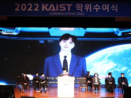
The 2022 commencement ceremony convened online on February 18 to celebrate and award degrees to the Class of 2022. The graduating class of 2022 included 663 PhDs, 1,383 Masters, and 695 Bachelors. The limited number of attendees included 86 graduate representatives and approximately 20 faculty members in senior leadership, as well as Korea’s Minister of Science and ICT Hyesook Lim. The ceremony was livestreamed on KAIST’s YouTube channel.
Valedictorian Ji-Young Lee from the Department of Physics received the Minister of Science and ICT’s Award. Yu-Jin Bang from the School of Business and Technology Management was the Awardee of the Chairman of the KAIST Board of Trustees and the KAIST Presidential Awardee was Jong-Hwan Lee from the Department of Mathematical Sciences.
KAIST conferred honorary doctorates to Honorary Chairman Jae-Chul Kim of Dongwon Group and Chairman Sung-Hwan Chang of Samsung Brush. Chairman Kim, whose donation funded the establishment of the Kim Jae-Chul Graduate School of AI, was awarded an honorary doctorate of science technology. Chairman Chang was awarded an honorary doctorate of business administration in recognition of his funding in the fields of medical science and engineering at KAIST.
This year’s undergraduate commencement speaker was Hye-Lin Park from the School of Computing. She has severe cerebral palsy and was the first student admitted to KAIST with a severe physical handicap.
“I loved mathematics and wanted to become a mathematician. When I learned programming in my second year, I was so mesmerized by it that I transferred to the School of Computing,” said Park, who plans to continue studying programming languages in graduate school at KAIST.
“I spent my entire life of 24 years in this beautiful wheelchair. Without the support and help of my parents, friends, and my special teachers who helped me move and study at the campus, I would not have made it this far,” said Park. For easier access to classrooms and facilities, KAIST started to remodel its facilities to make the entrance of buildings more wheelchair-friendly. Park made many suggestions to the Office of Student Life and the Facilities Management Office on how to ease mobility for handicapped people on campus. The physical education course that was required for graduation was also revised to stipulate exceptions.
Minister Lim stressed the role of young scientists and researchers in these times of digital transformation dominated by AI and the metaverse. She encouraged the graduates to carry out their dreams with warm hearts and challenging spirits.
KAIST President Kwang Hyung Lee also stressed the power of dreams, calling for graduates to dream big even in times of uncertainty.
“Humanity stands at an inflection point in history. The fourth industrial revolution and outbreak of Covid-19 have unfolded the grand global transformation. Although the future gives us new opportunities, it also comes with anxiety regarding the uncertainties ahead.”
“Dreams make your heart race and push us to live life to the fullest. Dreams will help you keep moving forward even in the face of adversity,” he said.
2022.02.18 View 11293
Commencement Ceremony Honors the Class of 2022
Third online commencement ceremony since the pandemic outbreak celebrates 2741 graduates
The 2022 commencement ceremony convened online on February 18 to celebrate and award degrees to the Class of 2022. The graduating class of 2022 included 663 PhDs, 1,383 Masters, and 695 Bachelors. The limited number of attendees included 86 graduate representatives and approximately 20 faculty members in senior leadership, as well as Korea’s Minister of Science and ICT Hyesook Lim. The ceremony was livestreamed on KAIST’s YouTube channel.
Valedictorian Ji-Young Lee from the Department of Physics received the Minister of Science and ICT’s Award. Yu-Jin Bang from the School of Business and Technology Management was the Awardee of the Chairman of the KAIST Board of Trustees and the KAIST Presidential Awardee was Jong-Hwan Lee from the Department of Mathematical Sciences.
KAIST conferred honorary doctorates to Honorary Chairman Jae-Chul Kim of Dongwon Group and Chairman Sung-Hwan Chang of Samsung Brush. Chairman Kim, whose donation funded the establishment of the Kim Jae-Chul Graduate School of AI, was awarded an honorary doctorate of science technology. Chairman Chang was awarded an honorary doctorate of business administration in recognition of his funding in the fields of medical science and engineering at KAIST.
This year’s undergraduate commencement speaker was Hye-Lin Park from the School of Computing. She has severe cerebral palsy and was the first student admitted to KAIST with a severe physical handicap.
“I loved mathematics and wanted to become a mathematician. When I learned programming in my second year, I was so mesmerized by it that I transferred to the School of Computing,” said Park, who plans to continue studying programming languages in graduate school at KAIST.
“I spent my entire life of 24 years in this beautiful wheelchair. Without the support and help of my parents, friends, and my special teachers who helped me move and study at the campus, I would not have made it this far,” said Park. For easier access to classrooms and facilities, KAIST started to remodel its facilities to make the entrance of buildings more wheelchair-friendly. Park made many suggestions to the Office of Student Life and the Facilities Management Office on how to ease mobility for handicapped people on campus. The physical education course that was required for graduation was also revised to stipulate exceptions.
Minister Lim stressed the role of young scientists and researchers in these times of digital transformation dominated by AI and the metaverse. She encouraged the graduates to carry out their dreams with warm hearts and challenging spirits.
KAIST President Kwang Hyung Lee also stressed the power of dreams, calling for graduates to dream big even in times of uncertainty.
“Humanity stands at an inflection point in history. The fourth industrial revolution and outbreak of Covid-19 have unfolded the grand global transformation. Although the future gives us new opportunities, it also comes with anxiety regarding the uncertainties ahead.”
“Dreams make your heart race and push us to live life to the fullest. Dreams will help you keep moving forward even in the face of adversity,” he said.
2022.02.18 View 11293 -
 Five Projects Ranked in the Top 100 for National R&D Excellence
Five KAIST research projects were selected as the 2021 Top 100 for National R&D Excellence by the Ministry of Science and ICT and the Korea Institute of Science & Technology Evaluation and Planning.
The five projects are:-The development of E. coli that proliferates with only formic acid and carbon dioxide by Distinguished Professor Sang Yup Lee from the Department of Chemical and Biomolecular Engineering
-An original reverse aging technology that restores an old human skin cell into a younger one by Professor Kwang-Hyun Cho from the Department of Bio and Brain Engineering-The development of next-generation high-efficiency perovskite-silicon tandem solar cells by Professor Byungha Shin from the Department of Materials Science and Engineering-Research on the effects of ultrafine dust in the atmosphere has on energy consumption by Professor Jiyong Eom from the School of Business and Technology Management-Research on a molecular trigger that controls the phase transformation of bio materials by Professor Myungchul Kim from the Department of Bio and Brain Engineering
Started in 2006, an Evaluation Committee composed of experts in industries, universities, and research institutes has made the preliminary selections of the most outstanding research projects based on their significance as a scientific and technological development and their socioeconomic effects. The finalists went through an open public evaluation. The final 100 studies are from six fields: 18 from mechanics & materials, 26 from biology & marine sciences, 19 from ICT & electronics, 10 from interdisciplinary research, and nine from natural science and infrastructure.
The selected 100 studies will receive a certificate and an award plaque from the minister of MSIT as well as additional points for business and institutional evaluations according to appropriate regulations, and the selected researchers will be strongly recommended as candidates for national meritorious awards.
In particular, to help the 100 selected research projects become more accessible for the general public, their main contents will be provided in a free e-book ‘The Top 100 for National R&D Excellence of 2021’ that will be available from online booksellers.
2022.02.17 View 11889
Five Projects Ranked in the Top 100 for National R&D Excellence
Five KAIST research projects were selected as the 2021 Top 100 for National R&D Excellence by the Ministry of Science and ICT and the Korea Institute of Science & Technology Evaluation and Planning.
The five projects are:-The development of E. coli that proliferates with only formic acid and carbon dioxide by Distinguished Professor Sang Yup Lee from the Department of Chemical and Biomolecular Engineering
-An original reverse aging technology that restores an old human skin cell into a younger one by Professor Kwang-Hyun Cho from the Department of Bio and Brain Engineering-The development of next-generation high-efficiency perovskite-silicon tandem solar cells by Professor Byungha Shin from the Department of Materials Science and Engineering-Research on the effects of ultrafine dust in the atmosphere has on energy consumption by Professor Jiyong Eom from the School of Business and Technology Management-Research on a molecular trigger that controls the phase transformation of bio materials by Professor Myungchul Kim from the Department of Bio and Brain Engineering
Started in 2006, an Evaluation Committee composed of experts in industries, universities, and research institutes has made the preliminary selections of the most outstanding research projects based on their significance as a scientific and technological development and their socioeconomic effects. The finalists went through an open public evaluation. The final 100 studies are from six fields: 18 from mechanics & materials, 26 from biology & marine sciences, 19 from ICT & electronics, 10 from interdisciplinary research, and nine from natural science and infrastructure.
The selected 100 studies will receive a certificate and an award plaque from the minister of MSIT as well as additional points for business and institutional evaluations according to appropriate regulations, and the selected researchers will be strongly recommended as candidates for national meritorious awards.
In particular, to help the 100 selected research projects become more accessible for the general public, their main contents will be provided in a free e-book ‘The Top 100 for National R&D Excellence of 2021’ that will be available from online booksellers.
2022.02.17 View 11889 -
 President Lee Presents Plans to Nurture Next-Generation Talents
President Lee stressed that nurturing medical scientists, semiconductor R&D personnel, startup entrepreneurs, and global innovators are key missions he will continue to pursue during a news conference
KAIST President Kwang Hyung Lee said that nurturing medical scientists, semiconductor R&D personnel, startup entrepreneurs, and global innovators are key missions he will continue to pursue during an online news conference marking the 1st anniversary of him becoming the president on February 15.
He said that nurturing physician-scientists is the most critical mission for KAIST to help the nation create a new growth engine. He said KAIST will help the nation drive the bio-industry and provide medical science resources for the nation’s health sector. To this end, he said that KAIST will open its Medical Science and Technology School by 2026.
“We plan to expand the current Graduate School of Medical Science and Engineering into a new Medical Science and Technology School that will focus entirely on a condensed MD-PhD course converging the fields of AI, bio, and physics,” he said.
The school aims to foster medical scientists whose research results will eventually be commercialized. He said that the university is now discussing revisions to related laws and regulations with the government and other universities.
To supply human resources to the semiconductor industry, President Lee said the university will add a campus in Pyongtaek City that will serve as an advanced convergence research hub in the field of next generation semiconductors in collaboration with Samsung Electronics and the city of Pyongtaek. The three-stage opening plan projected the final opening of the campus by 2036. During the first stage, which will be completed by 2026, it will construct the campus infrastructure in Pyongtaek city where Samsung Semiconductors runs two massive semiconductor complexes. By 2031, it plans to launch the open research platform including a future cities research center and future vehicles research center. The campus will open the global industrial collaboration cluster hub by 2036.
In the global arena, President Lee said he is working to open the New York campus with stakeholders in the United States. He announced the plan last December that was endorsed by New York-based entrepreneur Hee-Nam Bae, the chairman of Big Continent Inc. President Lee and Chairman Lee signed an MOU for the funding to open the campus in New York.
“We are discussing how to facilitate the plan and best accommodate the interests and potential of our students. Many ideas and plans are on the table and we think it will take longer than expected to finalize the plan,” explained President Lee.
However, he added that the basic idea is to offer art tech and health technology programs as well as an AI-based finance MBA at the New York campus, in addition to it serving as the startup accelerator of KAIST.
President Lee stressed the importance of technology commercialization when successfully launching KAIST Holdings last month to help spinoffs of KAIST labs accelerate their end results. He said that KAIST Holdings will build a virtuous supporting system to commercialize the technology startups coming from KAIST.
“We plan to list at least 10 KAIST startups on the KOSDAQ and two on the NASDAQ by 2031. KAIST Holdings also aims to nurture companies valued at a total of one billion KRW and earn 100 billion KRW in technology fees by 2031.
2022.02.17 View 12550
President Lee Presents Plans to Nurture Next-Generation Talents
President Lee stressed that nurturing medical scientists, semiconductor R&D personnel, startup entrepreneurs, and global innovators are key missions he will continue to pursue during a news conference
KAIST President Kwang Hyung Lee said that nurturing medical scientists, semiconductor R&D personnel, startup entrepreneurs, and global innovators are key missions he will continue to pursue during an online news conference marking the 1st anniversary of him becoming the president on February 15.
He said that nurturing physician-scientists is the most critical mission for KAIST to help the nation create a new growth engine. He said KAIST will help the nation drive the bio-industry and provide medical science resources for the nation’s health sector. To this end, he said that KAIST will open its Medical Science and Technology School by 2026.
“We plan to expand the current Graduate School of Medical Science and Engineering into a new Medical Science and Technology School that will focus entirely on a condensed MD-PhD course converging the fields of AI, bio, and physics,” he said.
The school aims to foster medical scientists whose research results will eventually be commercialized. He said that the university is now discussing revisions to related laws and regulations with the government and other universities.
To supply human resources to the semiconductor industry, President Lee said the university will add a campus in Pyongtaek City that will serve as an advanced convergence research hub in the field of next generation semiconductors in collaboration with Samsung Electronics and the city of Pyongtaek. The three-stage opening plan projected the final opening of the campus by 2036. During the first stage, which will be completed by 2026, it will construct the campus infrastructure in Pyongtaek city where Samsung Semiconductors runs two massive semiconductor complexes. By 2031, it plans to launch the open research platform including a future cities research center and future vehicles research center. The campus will open the global industrial collaboration cluster hub by 2036.
In the global arena, President Lee said he is working to open the New York campus with stakeholders in the United States. He announced the plan last December that was endorsed by New York-based entrepreneur Hee-Nam Bae, the chairman of Big Continent Inc. President Lee and Chairman Lee signed an MOU for the funding to open the campus in New York.
“We are discussing how to facilitate the plan and best accommodate the interests and potential of our students. Many ideas and plans are on the table and we think it will take longer than expected to finalize the plan,” explained President Lee.
However, he added that the basic idea is to offer art tech and health technology programs as well as an AI-based finance MBA at the New York campus, in addition to it serving as the startup accelerator of KAIST.
President Lee stressed the importance of technology commercialization when successfully launching KAIST Holdings last month to help spinoffs of KAIST labs accelerate their end results. He said that KAIST Holdings will build a virtuous supporting system to commercialize the technology startups coming from KAIST.
“We plan to list at least 10 KAIST startups on the KOSDAQ and two on the NASDAQ by 2031. KAIST Holdings also aims to nurture companies valued at a total of one billion KRW and earn 100 billion KRW in technology fees by 2031.
2022.02.17 View 12550 -
 Perigee-KAIST Rocket Research Center Launches Scientific Rocket
Undergraduate startup Perigree Aerospace develops suborbital rocket called Blue Whale 0.1
On December 29, Perigee Aerospace, an undergraduate startup, launched a test rocket with a length of 3.2 m, a diameter of 19 cm, and a weight of 51 kg, using ethanol and liquid oxygen as fuel. The launch took place off Jeju Island. It was aimed at building experience and checking the combustion of a liquid propulsion engine and the performance of pre-set flight and trajectory, communication, and navigation devices. It was also one of the projects marking the 50th anniversary of KAIST in 2021.
However, after flying for several seconds, the rocket lost its track due to a gust of wind that activated the rocket’s automatic flight suspension system. "At the moment the rocket took off, there was a much stronger gust than expected," Dong-Yoon Shin, CEO of Perigee said. "The wind sent it flying off course and the automatic flight suspension system stopped its engine." However, Shin was not disappointed, saying the launch, which was conducted in collaboration with Perigee-KAIST Rocket Research Center provided a good experience.
"Some people say that Blue Whale 0.1 is like a toy because of its small size. Of course, it's much smaller than the rockets I’ve dreamed of, but like other rockets, it has all the technology needed for launch," said Shin, who established his company in 2018 as a KAIST aerospace engineering student to develop small liquid-propellant orbital rockets. Perigee Aerospace aims to develop the world’s lightest launch vehicle using high-powered engines, with a goal of leading the global market for small launch vehicles in the new space generation.
Perigee-KAIST Rocket Research Center was founded in 2019 for the research and development of rocket propellants and has been testing the combustion of rocket engines of various sizes in their liquid propellant rocket combustion lab located on the KAIST Munji Campus.
The research center initiated the 50th anniversary rocket launch project in late April of last year, finished the examination of their preliminary design in late May, and secured a tentative launching site through the KAIST-Jejudo agreement in early July.
The ethanol engine combustion was tested in late July, and an examination meeting regarding the detailed design that took place in late August was followed by two months of static firing tests of the assembled rocket in October and November.
This was a very meaningful trial in which a domestic private enterprise founded by a college student collaborated with a university to successfully develop and launch a technically challenging liquid propellant rocket.
Shin's near-term goal is to launch a two-stage orbital rocket that uses liquid methane as fuel and weighs 1.8 tons. To secure competitiveness in the small projectile market, KAIST and Perigee Aerospace have set up a joint research center to test various rocket engine sizes and develop the world's lightest projectile using a high-performance engine.
Professor Jae-Hung Han, head of the Department of Aerospace Engineering, said, “The scientific rocket system secured through the launch of the celebratory rocket will be utilized for design and system-oriented education, and for carrying out various scientific missions.” He added, “It is very rare both domestically and globally that a scientific rocket designed by the initiatives of a department should be incorporated as part of a regular aerospace system design curriculum. This will be an exemplary case we can boast about to the rest of the world.”
Perigee Aerospace will improve the technology they have developed through the course of this project to develop subminiature vehicles they may use to launch small satellites into the low Earth orbit.
Shin said, “I am happy just with the fact that we have participated in a rocket project to celebrate the 50th anniversary of KAIST, and I would like to thank the engineers at my company and members of the KAIST Department of Aerospace Engineering.” He added, “I’m looking forward to the day that we develop a space launch vehicle that can deliver satellites even higher.”
2022.01.14 View 10119
Perigee-KAIST Rocket Research Center Launches Scientific Rocket
Undergraduate startup Perigree Aerospace develops suborbital rocket called Blue Whale 0.1
On December 29, Perigee Aerospace, an undergraduate startup, launched a test rocket with a length of 3.2 m, a diameter of 19 cm, and a weight of 51 kg, using ethanol and liquid oxygen as fuel. The launch took place off Jeju Island. It was aimed at building experience and checking the combustion of a liquid propulsion engine and the performance of pre-set flight and trajectory, communication, and navigation devices. It was also one of the projects marking the 50th anniversary of KAIST in 2021.
However, after flying for several seconds, the rocket lost its track due to a gust of wind that activated the rocket’s automatic flight suspension system. "At the moment the rocket took off, there was a much stronger gust than expected," Dong-Yoon Shin, CEO of Perigee said. "The wind sent it flying off course and the automatic flight suspension system stopped its engine." However, Shin was not disappointed, saying the launch, which was conducted in collaboration with Perigee-KAIST Rocket Research Center provided a good experience.
"Some people say that Blue Whale 0.1 is like a toy because of its small size. Of course, it's much smaller than the rockets I’ve dreamed of, but like other rockets, it has all the technology needed for launch," said Shin, who established his company in 2018 as a KAIST aerospace engineering student to develop small liquid-propellant orbital rockets. Perigee Aerospace aims to develop the world’s lightest launch vehicle using high-powered engines, with a goal of leading the global market for small launch vehicles in the new space generation.
Perigee-KAIST Rocket Research Center was founded in 2019 for the research and development of rocket propellants and has been testing the combustion of rocket engines of various sizes in their liquid propellant rocket combustion lab located on the KAIST Munji Campus.
The research center initiated the 50th anniversary rocket launch project in late April of last year, finished the examination of their preliminary design in late May, and secured a tentative launching site through the KAIST-Jejudo agreement in early July.
The ethanol engine combustion was tested in late July, and an examination meeting regarding the detailed design that took place in late August was followed by two months of static firing tests of the assembled rocket in October and November.
This was a very meaningful trial in which a domestic private enterprise founded by a college student collaborated with a university to successfully develop and launch a technically challenging liquid propellant rocket.
Shin's near-term goal is to launch a two-stage orbital rocket that uses liquid methane as fuel and weighs 1.8 tons. To secure competitiveness in the small projectile market, KAIST and Perigee Aerospace have set up a joint research center to test various rocket engine sizes and develop the world's lightest projectile using a high-performance engine.
Professor Jae-Hung Han, head of the Department of Aerospace Engineering, said, “The scientific rocket system secured through the launch of the celebratory rocket will be utilized for design and system-oriented education, and for carrying out various scientific missions.” He added, “It is very rare both domestically and globally that a scientific rocket designed by the initiatives of a department should be incorporated as part of a regular aerospace system design curriculum. This will be an exemplary case we can boast about to the rest of the world.”
Perigee Aerospace will improve the technology they have developed through the course of this project to develop subminiature vehicles they may use to launch small satellites into the low Earth orbit.
Shin said, “I am happy just with the fact that we have participated in a rocket project to celebrate the 50th anniversary of KAIST, and I would like to thank the engineers at my company and members of the KAIST Department of Aerospace Engineering.” He added, “I’m looking forward to the day that we develop a space launch vehicle that can deliver satellites even higher.”
2022.01.14 View 10119